| Cardiac | |
|---|---|
| GI | |
| Bone | |
| GU | |
| Neuro | |
| Peds | |
| Faculty | |
| Student | |
| Quizzes | |
| Image DDX | |
| Museum | |
| Mobile | |
| |
Misc |
| Videocasts | |
| Signs | |
Learning
Radiology:
Recognizing
the Basics
Available
on the Kindle
and IPad
LearningRadiology Imaging Signs
on Twitter
![]()
Follow us on
What is the most likely diagnosis?
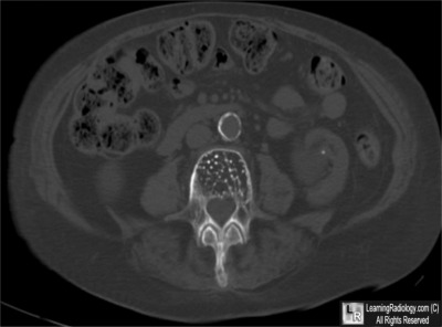
- 67 year-old complaining of low back pain

Close-up of Lower Lumbar Spine
Lateral Radiograph
- Paget Disease
- Renal Osteodystrophy
- Hemangioma
- Metastases
- Multiple Myeloma
Additional image - Axial CT of Spine
Answer:
3. Hemangioma
More (Click Discussion Tab)
Hemangioma
General Considerations
- Benign
- Most often located in lower thoracic, upper lumbar spine
- Skull is second most common location (spoke-wheel appearance)
- Mostly asymptomatic
- More frequent in females
- Peak incidence in 40’s
- Multiple in up to 1/3 of cases
- Most often occur in the medullary cavity of bone
- Microscopically, there is hamartomatous proliferation of vascular tissue
- Classified as to cavernous, capillary, arteriovenous and venous
- Spine hemangiomas are usually capillary type; skull are cavernous
MORE . . .
.
This Week
67 year-old complaining of low back pain |
Some of the fundamentals of interpreting chest images |
The top diagnostic imaging diagnoses that all medical students should recognize according to the Alliance of Medical Student Educators in Radiology |
Recognizing normal and key abnormal intestinal gas patterns, free air and abdominal calcifications |
Recognizing the parameters that define a good chest x-ray; avoiding common pitfalls |
How to recognize the most common arthritides |
LearningRadiology
Named Magazine's
"25 Most Influential"

See Article on LearningRadiology
in August, 2010
RSNA News
| LearningRadiology.com |
is an award-winning educational website aimed primarily at medical students and radiology residents-in-training, containing lectures, handouts, images, Cases of the Week, archives of cases, quizzes, flashcards of differential diagnoses and “most commons” lists, primarily in the areas of chest, GI, GU cardiac, bone and neuroradiology. |


