
| Cardiac | |
|---|---|
| GI | |
| Bone | |
| GU | |
| Neuro | |
| Peds | |
| Faculty | |
| Student | |
| Quizzes | |
| Image DDX | |
| Mobile | |
| |
Misc |
| Videocasts |
 |
![]() Case of the Week 346
Case of the Week 346 ![]()
What's the most likely diagnosis?
- 41 year-old with left lower quadrant pain
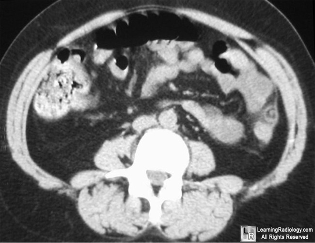
Axial CT of Lower Abdomen
- Colonic polyp
- Shock bowel
- Pneumoperitoneum
- Epiploic appendagitis
- Absence of the kidneys
Additional Image - Another axial CT slice of lower abdomen
![]()
Additional Image
.
.
Axial CT of Lower Abdomen
.
![]()
Answer:
.
4. Epiploic Appendagitis
.
.
More (Click Discussion Tab)
Epiploic Appendagitis
General Considerations
- Uncommon cause of acute abdominal pain
- Occurs most often in men in 4th and 5th decade
- Inflammation of epiploic appendages (appendices)
- Peritoneal outpouchings originating from serosal surface of colon
- Contain blood vessels and fat
- Diagnosis requires cross-sectional imaging
- Inflammation may be caused by venous occlusion
- “Secondary” epiploic appendagitis is caused by inflammation of an adjacent structure
.
This Week
The 2nd of a two-part video podcast reviews the causes and appearances of soft tissue calcifications associated with arthritis, tumors, trauma and more; there is an expanded mini-quiz at the end |
A trim mobile version for your PDA or internet-enabled smartphone with handy lists of differential diagnoses, most commons and more
|
Key points on recognizing the most common fractures and dislocations |
Basic CT imaging of the brain focusing on the findings of cerebrovascular accidents |
The top diagnostic imaging diagnoses that all medical students should recognize according to the Alliance of Medical Student Educators in Radiology |
Recognizing normal and key abnormal intestinal gas patterns, free air and abdominal calcifications |
Some of the fundamentals of interpreting chest images |
UPDATED ![]()
| LearningRadiology.com |
is an award-winning educational website aimed primarily at medical students and radiology residents-in-training, containing lectures, handouts, images, Cases of the Week, archives of cases, quizzes, flashcards of differential diagnoses and “most commons” lists, primarily in the areas of chest, GI, GU cardiac, bone and neuroradiology. |