|
|
Umbilical Venous Catheter in Left Ventricle
For more on Umbilical Venous and Arterial Catheters, including their normal positioning, uses and examples of aberrant positioning, click on this link
Normal Positioning
-
From the umbilicus, umbilical vein passes cephalad, slightly to right
-
Joins the left branch of the portal vein
-
The ductus venosus arises from the point where the UV joins the left portal vein
-
Ductus venosus enters IVC
-
UVC should be just above diaphragm where IVC enters RA (T8-T9)
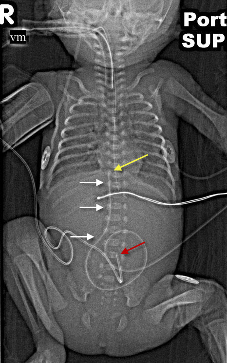
Umbilical Venous Catheter (UVC) at T9. There is an UVC that ascends slightly to the right and over the spine (white arrows) to the level of T9 (yellow arrow). The umbilical arterial catheter (UAC) is at L4 (red arrow).
From Auckland District Health Board
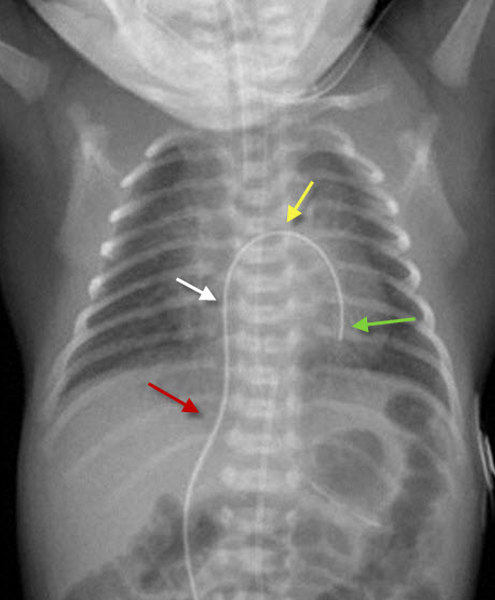
Umbilical Venous Catheter (UVC) in region of Left Ventricle. An umbilical venous catheter ascends to the right of the spine (red arrow). It continue into the inferior vena cava and right atrium (white arrow). It then passes through a patent foramen ovale into the left side of the heart (yellow arrow) and finally into the left ventricle (green arrow).
|
|
|
