
| Cardiac | |
|---|---|
| GI | |
| Bone | |
| GU | |
| Neuro | |
| Peds | |
| Faculty | |
| Student | |
| Quizzes | |
| Image DDX | |
| Museum | |
| Mobile | |
| |
Misc |
| Videocasts | |
| Signs | |
 |
Learning
Radiology:
Recognizing
the Basics
Available
on the Kindle
and IPad
LearningRadiology Imaging Signs
on Twitter
![]()
Follow us on
![]()
![]() Case of the Week 560
Case of the Week 560 ![]()
What is the most likely diagnosis?
- 63 year-old male with history of arthritis

Frontal Chest Radiograph
- Histiocytosis X
- Septic Emboli
- Erosive Osteoarthritis
- Rheumatoid Nodules
- Reactive Arthritis
Additional Images - Axial CT images of the chest
![]()
Additional Images
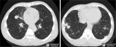
Axial CT images of the chest
![]()
Answer:
4. Rheumatoid Nodules
More (Click Discussion Tab)
Rheumatoid Nodules
General Considerations
- Rare
- Immune-mediated granulomas frequently with necrotic centers
- They are almost always associated with long, standing active rheumatoid arthritis
- More frequent in males with high titers for rheumatoid factor
- Also more frequent in smokers and those who already have subcutaneous nodules
MORE . . .
.
This Week
63 year-old male with history of arthritis |
Some of the fundamentals of interpreting chest images |
The top diagnostic imaging diagnoses that all medical students should recognize according to the Alliance of Medical Student Educators in Radiology |
Recognizing normal and key abnormal intestinal gas patterns, free air and abdominal calcifications |
Recognizing the parameters that define a good chest x-ray; avoiding common pitfalls |
How to recognize the most common arthritides |
LearningRadiology
Named Magazine's
"25 Most Influential"
![]()
See Article on LearningRadiology
in August, 2010
RSNA News
| LearningRadiology.com |
is an award-winning educational website aimed primarily at medical students and radiology residents-in-training, containing lectures, handouts, images, Cases of the Week, archives of cases, quizzes, flashcards of differential diagnoses and “most commons” lists, primarily in the areas of chest, GI, GU cardiac, bone and neuroradiology. |
![]()