|
|
Urinary Bladder Hernias
General Considerations
- About 1-3% of inguinal hernias involve urinary bladder
- Femoral hernias are more common in women, inguinal in men
- More often on right side
- Small portion to almost all of bladder may herniate
- Damage during a herniorrhaphy can occur making imaging an important study prior to surgery
- Contributing factors to developing a bladder hernia include
- Chronic outlet obstruction and prolonged bladder distension
- Loss of bladder tine
- Pericystitis
- Obesity
- Space-occupying pelvic masses
Clinical Findings
- Most are asymptomatic
- Dysuria
- Frequency
- Urgency
- Nocturia and hematuria
- Inguinal “mass” which may empty after voiding
Imaging Findings
- Findings are the same regardless of the imaging modality
- CT is best for showing overall anatomy of the hernia
- Wide-mouthed, rounded protrusion of the bladder inferiorly, anteriorly and laterally
- Asymmetric bladder
- The hernia may be less evident or not seen on supine imaging; it may become visible only after voiding
Differential Diagnosis
- In infants, small protrusions from the base of the bladder laterally are called “bladder ears” and are considered normal
Classification of Bladder Hernias |
Type
|
Remarks
|
Paraperitoneal |
Most Frequent; extraperitoneal portion of hernia lies along medial wall of sac |
Intraperitoneal |
Completely
covered by peritoneum |
Extraperitoneal |
Bladder herniates without
any relation with the peritoneum |
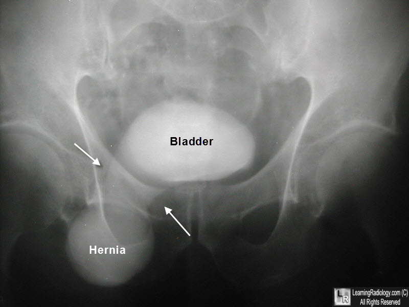
Bladder Hernia. There is contrast in the urinary bladder (bladder). A smooth protrusion points inferiorly through the right inguinal canal (white arrows) into the right groin (hernia).
Imaging of Urinary Bladder Hernias. LE Bacigalupo, , M Bertolotto,, F Barbiera,, P Pavlica, R Lagalla , RS. Pozzi Mucelli and LE. Derchi. AJR 2005;184:546–551
|
|
|
