|
|
Bear Paw Sign
General Considerations
- The appearance of the kidney on CT, usually associated with xanthogranulomatous pyelonephritis
- Features calcifications in a non-functioning kidney that has multiple collections of hyperattenuating masses which resemble a pattern of hydronephrosis
- There may be enhancement of the margins of these masses after contrast administration.
- This appearance on CT scans has been described as the bear paw sign
- It has also been described in congenital uretero-pelvic junction obstruction
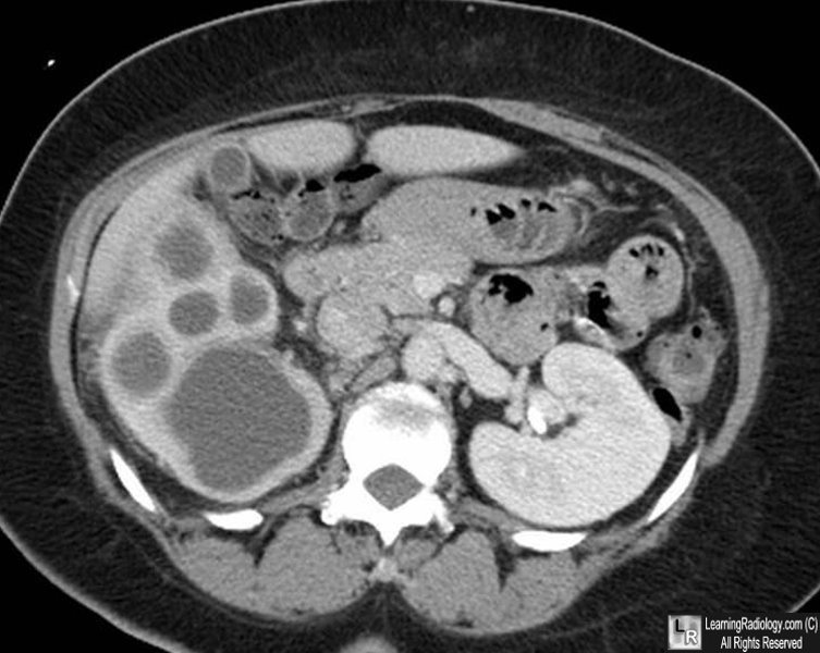
Bear Paw Sign. Axial, enhanced CT of the right kidney displays a pattern of large "hydronephrotic" collections in the regions of the calyces in the shape of a bear's paw. The renal pelvis is not dilated. The sign is associated with xanthogranulomatous pyelonephritis.
Evolving concepts in the diagnosis of xanthogranulomatous pyelonephritis. Parker MD, Clark RL Urol Radiol 1989; 11:7-15.
Classic Signs in Uroradiology. RB Dyer, MY Chen, and R. Zagoria,. RadioGraphics, October 2004, Volume 24, Issue suppl_1
|
|
|
