|
|
Calcified Thyroid Adenoma
General Considerations
- Thyroid adenomas are benign neoplastic growths contained within a capsule
- They are characteristically enclosed by a discrete capsule or a thin rim of compressed surrounding thyroid tissue
- Adenomas may become partially cystic, probably through necrosis of a portion of the growth
- Cyst formation is very common in colloid nodules, a type of adenoma
- Patients with the hereditable multiple endocrine neoplasia syndrome (MEN), type I, may have thyroid adenomas, parathyroid adenomas, islet cell tumors, and adrenal tumors
Clinical Findings
- Typically, they are asymptomatic
- It is unusual for these growths to attain a size capable of producing symptoms such as dysphagia or stridor
- Hemorrhage into an adenoma can result in acute pain
Imaging Findings
- Peripheral, rim calcifications suggests a benign lesion, especially a degenerated thyroid adenoma
- Plain films may only show an enlarged thyroid displacing the trachea or calcification within a mass
- Ultrasound is the initial study of choice for evaluating a thyroid mass
- Most cystic lesions are benign
- Malignant nodules are mostly solid and hypoechoic, occasionally with a small punctate calcification
- CT and MRI are used mostly for identifying lymphadenopathy or metastases
- About 10% of thyroid adenomas are “hot” on thyroid scans
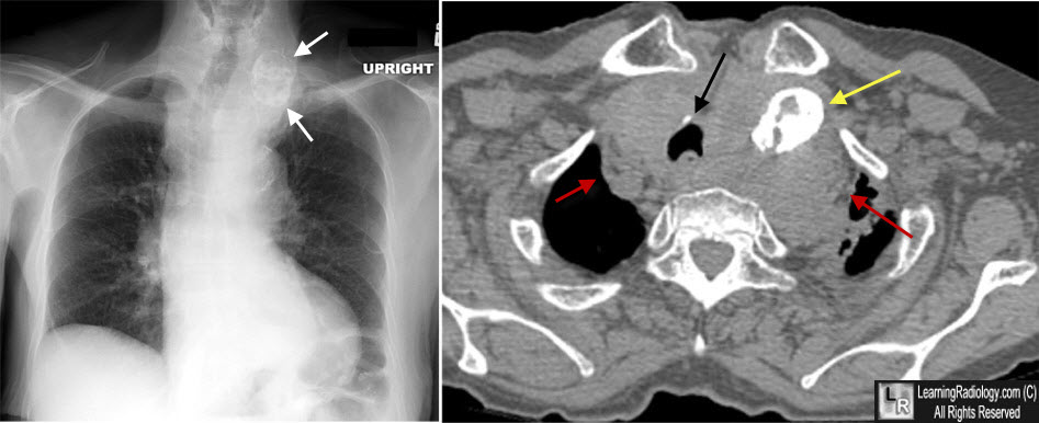
Calcified Thyroid Adenoma. The frontal chest radiograph shows a calcified structure in the left side of the lower neck, upper chest which has both peripheral and internal calcifications (white arrows). The CT scan on the same patient shows a large substernal thyroid (red arrows) displacing the trachea to the right (black arrow) and containing a large degenerated thyroid adenoma (yellow arrow).
Thyroid Nodules. LJ DeGroot and F Pacini. Thyroid Disease Manager. March 10, 2012.
|
|
|
