|
|
Swyer James Syndrome
MacLeod Syndrome
General Considerations
- Rare
- Unilateral hyperlucent lung
- Acquired post-infectious obliterative bronchiolitis occurring in childhood
- Usually post-viral
- Changes occur months to years after causative infection
- Involved lung is smaller than the opposite side
Clinical Findings
- May be asymptomatic
- Recurrent respiratory infections
- Productive cough
- Shortness of breath
- Dyspnea on exertion
- Hemoptysis
Imaging Findings
- Hyperlucency of all or part of a lung
- Diminished arterial flow with hypogenesis of the pulmonary arteries
- Overexpansion of the contralateral lung
- Bronchiectasis may be present
- On CT, bronchi are pruned and there is air-trapping
- There is underdevelopment of peripheral pulmonary vessels
- On nuclear imaging, there is decreased perfusion of the affected lung and delayed entry and washout on ventilation scans
Differential Diagnosis
- Hypoplastic lung syndrome
- Bronchiolitis obliterans – more diffuse than Swyer James
Treatment
- Early control of lung infections
- Flu and pneumococcal vaccinations
- If needed, resection of the affected lung
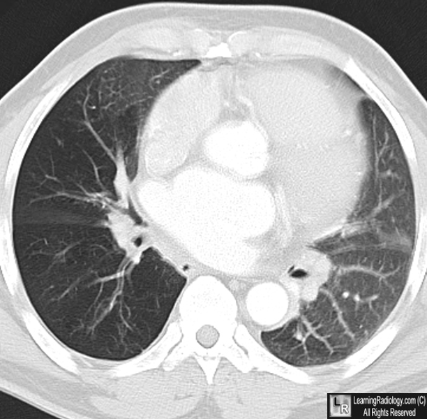
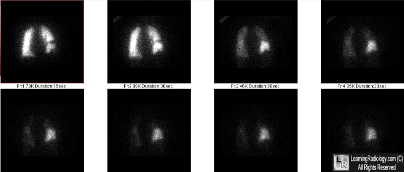
Swyer James Syndrome. (Top) Axial enhanced CT scan shows decreased vasculature in the right lower lobe, compared to the rest of the lungs. The peripheral pulmonary vasculature, in particular, is hypoplastic. (Bottom) A ventilation nuclear medicine scan is shown. The right lung is on your right. Note how the right lower lobe retains the tracer long after the remainder of the lung activity has washed out, because of air-trapping. The images are taken serially with the earliest at the top left and the latest at the bottom right.
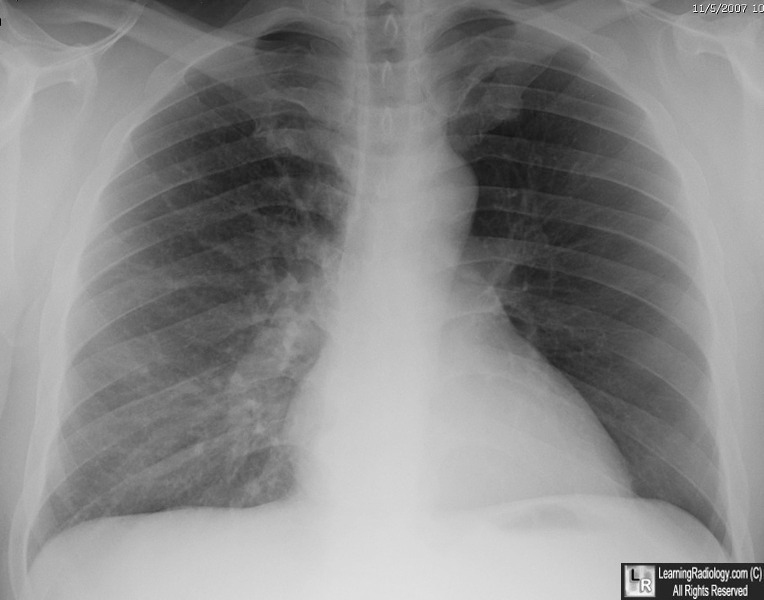
Swyer James Syndrome. Another patient with Swyer-James Syndrome, this time of the left lung. Notice the left hemithorax is more lucent than the right. On closer examination, note how many fewer pulmonary blood vessels there are on the left than the right. The left hilum is also smaller then the right.
Swyer-James Syndrome: CT Findings in Eight Patients. JD Godwin, PA Dietnch, JA. Verschakelen, WA Henderson, Jr. AJR 158:1211-1215, June 1992
|
|
|
