|
|
Superior Vena Caval Obstruction
Superior Vena Caval Syndrome
General Considerations
- Obstruction of flow in the superior vena cava due to:
- Most often due to thoracic malignancy, especially small-cell carcinoma
- Large-cell type of non-Hodgkin lymphoma
- Intravascular thrombosis indwelling catheters
- Mediastinal fibrosis
- Infections such as TB and histoplasmosis
- May produce superior vena caval syndrome
- Obstruction of the SVC results in one or more collateral pathways (60% or more narrowing)
- The azygous venous system, which includes the azygos vein, the hemiazygos vein, and the connecting intercostal veins
- The internal mammary venous system plus tributaries and secondary communications to the superior and inferior epigastric veins
- The long thoracic venous system, with its connections to the femoral veins and vertebral veins, provides the third and fourth collateral routes.
Clinical Findings
- Symptoms depend, in large measure, on how quickly the obstruction develops and the efficiency of the collaterals that are able to form
- Dyspnea is the most common symptom
- Facial swelling
- Cough
- Arm swelling
- Hoarseness
- Stridor
Imaging Findings
- Chest radiography
- Mediastinal or right upper lobe mass
- CT is the study of choice
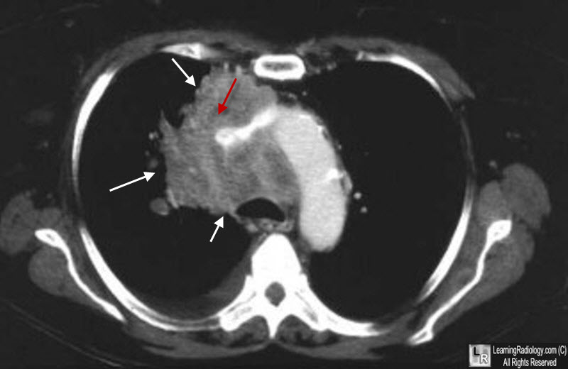
- Mediastinal mass
- Narrowing or complete obstruction of the SVC
- Collateral flow
- Venography is the most conclusive study
Treatment
- Relieve symptoms with oxygen, diuretics, elevation of the head
- Treat the primary malignancy, if present
- Thrombolysis, if indicated
Prognosis
- Depends on the underlying cause of the obstruction

Superior vena Caval Obstruction. There is a mediastinal soft tissue mass (white arrows) that is completely obstructing the superior vena cava, the location of which is marked by a red arrow.
Superior Vena Cava Syndrome Treatment and Management. TA Nickloes, AM Kallab, and LO Mack. eMedicine.
|
|
|