|
|
Silhouette Sign
General Considerations
- If two objects of the same radiographic density touch each other, the edge or margin between them disappears and it will be impossible to tell where one object begins and the other ends
- Conventional radiography is limited to demonstrating five basic densities
- Air, which appears the blackest on a radiograph
- Fat
- Soft tissue or fluid (because both soft tissue and fluid appear the same on conventional radiographs explains, for example, why you can’t differentiate between heart muscle and the blood inside of the heart on a chest radiograph)
- Calcium (usually contained within bones)
- Metal, which appears the whitest on a radiograph
- Objects of metal density do not appear normally
- Radiologic contrast media and foreign bodies are examples of metal densities artificially placed in the body
- The silhouette sign is valuable not only in the chest but as an aid in the analysis of imaging studies throughout the body
- Remember: two conditions must exist in order to have a positive silhouette sign
- The two objects in question must be in contact with each other
- They must be the same radiographic density (e.g. fat and fat, soft tissue and soft tissue, etc.)
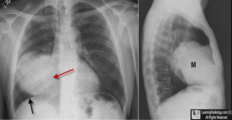
Silhouette Sign, Right Middle Lobe Mass. On the frontal image, there is a large mass in the right lower lung field. We note it is "silhouetting" the right heart border (red arrow) which is no longer seen as a distinct edge. It is not silhouetting the right hemidiaphragm (black arrow). The mass is therefore (1) touching the right heart border and is anterior and (2) the mass is soft tissue or fluid density. The lateral view shows the mass (M) is in the right middle lobe. It was a large bronchogenic carcinoma.
|
|
|
