|
|
Pneumonia - Lingula of Left Upper Lobe
General Considerations
- It is always best to localize disease on conventional radiographs using two views taken at 90° to each other (orthogonal views) like a frontal and lateral chest radiograph
- Sometimes, only a frontal radiograph may be available, as in critically ill or debilitated patients who require a portable bedside examination
- Nevertheless, it is still frequently possible to localize the pneumonia using only the frontal radiograph by analyzing which structure’s edges are obscured by the disease
- Silhouette sign
- If two objects of the same radiographic density touch each other, then the edge between them disappears
- The silhouette sign is valuable in localizing and identifying tissue types throughout the body, not just the chest
- You can utilize the silhouette sign to localize a pneumonia, even if only a frontal projection is available
Using the Silhouette Sign on the Frontal Chest Radiograph |
If this structure is no longer visible |
Then the disease is located in the |
Ascending aorta |
Right upper lobe |
Right heart border |
Right middle lobe |
Right hemidiaphragm |
Right lower lobe |
Descending aorta |
Left upper or lower lobe |
Left heart border |
Lingula of left upper lobe |
Left hemidiaphragm |
Left lower lobe |
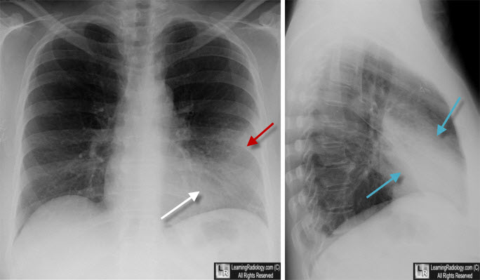
Lingular Pneumonia. The frontal view shows an airspace density in the left lower lung field (red arrow) which is silhouetting the left heart border (white arrow). The lateral view confirms the pneumonia is anterior, in the region of the lingula (blue arrows)
|
|
|
