|
|
Azygos Lobe
General Considerations
- Found as an anatomic variant in about 1% of people, the azygos lobe forms when the posterior cardinal vein, instead of remaining along the apex of the lung, penetrates the upper medial portion of the apical segment of the right upper lobe
- Its demarcating fissure contains both visceral and parietal pleural layers
- It has no bronchus so is not a true accessory lobe
- The size of the lobe is variable
Clinical Findings
Imaging Findings
- A thin, curvilinear density is seen in the upper right lung, convex towards the chest wall
- At its base is the azygos vein which can be seen as a teardrop-shaped structure
- In those with an azygos fissure, the azygos vein will no longer be visible at its normal position at the junction of the trachea and right main bronchus
Differential Diagnosis
- It should not be mistaken for an abnormality like a pneumothorax or bulla
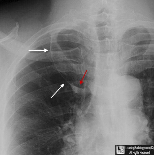
Azygos Lobe. The azygos fissure (white arrows) demarcates the lateral boundary of the azygos lobe, part of the right upper lobe. The tear-shaper azygos vein is seen at the base of the fissure (red arrow).
Imaging of the Azygos Lobe: Normal Anatomy and Variations. Josep Mata, Jose Caceres, Xavier Alegret, Pilar Coscojuela, and Jose Angel De Marcos.. AJR 156:931-937, May 1991
|
|
|
