|
|
Left Ventricular Thrombus
General Considerations
- Frequent complication of acute myocardial infarction (AMI) and dilated cardiomyopathy
- Most often seen in those with large anterior infarcts with ST-elevation (STEMI) apicoanterior aneurysm formation
- Significance lies in its ability to arterially embolize
- Contributing factors include
- Regional-wall akinesia or dyskinesia
- Endocardial inflammation providing a thrombogenic surface
- Hypercoagulable state
Clinical Findings
- Most frequent clinical presentation is the occurrence of a stroke after AMI, usually within the first 10 days
Imaging Findings
- Transthoracic echocardiography is the imaging modality of first choice
- CT’s specificity and sensitivity in detecting LV mural thrombus are similar to 2-D transthoracic echocardiography
- CT can detect new or organized thrombi as small as 2-3 cm in diameter
- Requires intravenous contrast administration
- Diagnosis is aided by the lack of enhancement of thrombi versus cardiac tumors
- Delayed enhancement cardiac MRI is very useful in identifying left ventricular aneurysms
- Gadolinium may be used to help differentiate myocardium from thrombus
- The avascular LVT is viewed as an absence of contrast uptake adjacent to hyper-enhanced, scarred myocardium
Differential Diagnosis
- Myxoma – usually occurs on the interatrial septum and projects into left atrium
Treatment
- Anticoagulant therapy may substantially decrease the rate of embolic events by 33% compared with no anticoagulation
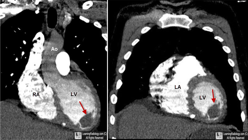
Left Ventricular Thrombus, CT. Two, reformatted, gated-CT images of the heart show a filling defect (red arrow) in the apex of the left ventricle (LV). LA=left atrium, RA=right atrium, Ao=aorta.
Left ventricular thrombus. Peter J. Stokman, Charn S. Nandra, Richard W. Asinger. Current Treatment Options in Cardiovascular Medicine. December 2001, Volume 3, Issue 6, pp 515-521
|
|
|
