|
|
Central Venous Catheter in Azygos Vein
General Considerations
- The azygos vein carries blood coursing mostly along the right side of the vertebral column in the posterior mediastinum towards the superior vena cava (SVC), joining the SVC at the level of T5
- It connects the systems draining into the SVC with the systems draining into the inferior vena cava (IVC)
- Thus it serves as a collateral pathway back to the right atrium if either the SVC or IVC is obstructed
- In approximately 2% of the population, the arch of the azygos vein which courses over the right main bronchus, can be part of a pleural septum producing the azygos lobe, which is separate from the right upper lobe
- It is an unpaired vein. Its counterparts on the left side are the hemiazygos and accessory hemiazygos veins
- Cannulation of the azygos vein is more likely to occur from a left-sided approach, but is rare overall (1.2%) of CVC insertions
- It also may be more common in patients with congestive heart failure, as the vein may be dilated
- The oxygen concentration of blood in the azygos vein is higher than the SVC
Imaging Findings
- The azygos vein may be seen on normal chest radiographs at the right junction of the trachea and the right main bronchus
- It may appear larger on supine studies than on upright images and may enlarge if it is carrying collateral flow from, for example, an obstructed inferior vena cava
- After central venous catheter (CVC) insertion, the tip may be seen on the frontal view abruptly looping upward and posteriorly in the right superior mediastinum
- On the lateral view, the CVC placed in the azygos will course posteriorly
Prognosis
- Perforation is very rare
- Fistulae between catheters placed in the azygos vein and adjacent airways has been described but is extremely rare
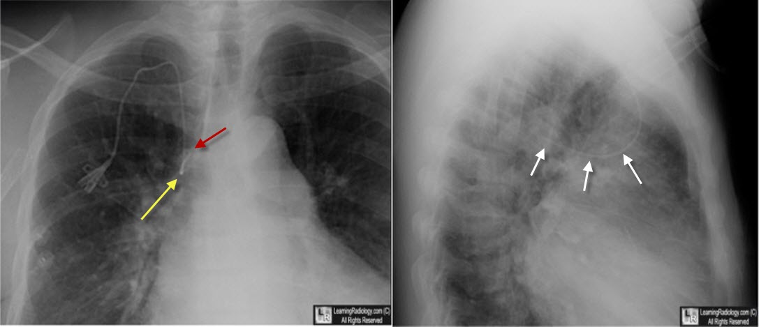
Central Venous Catheter in Azygos Vein. Image on the left shows a right-sided central venous catheter (CVC) directed first downward in the superior vena cava ((SVC) (yellow arrow) but then looping on itself upwards and to the left (red arrow). The lateral view shows the catheter directed posteriorly (white arrows) in the azygos arch.
 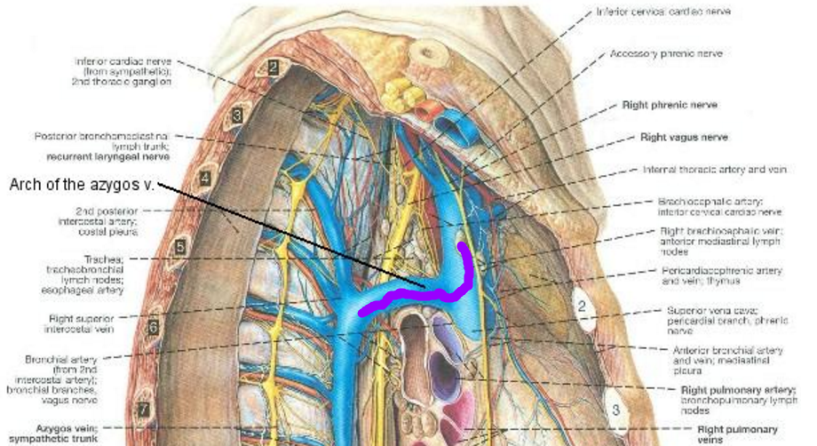
Click on images to enlarge. On left, the red line shows the rote of a left-sided CVC into the azygos in the frontal projection (from Netter Elsevier Images). On the right, the purple line shows the posterior projection of the azygos arch in the lateral projection. (from the University of Cincinnati)
The azygos vein pathway: an overview from anatomical variations to pathological changes. S Piciucchi, D Barone, S Sanna,
A Dubini, LR Goodman, D Oboldi, M Bertocco, C Ciccotosto, G Gavelli, A Carloni, and V Poletti. Insights Imaging. 2014 Oct; 5(5): 619–628.
|
|
|
