|
|
Spondylolysis
General Considerations
- The pars interarticularis is the bridge of bone that connects the superior and inferior articulating facets. They are paired, right and left
- Lumbar spondylolysis is a defect in the pars that may affect one or both sides and involve one or more vertebral bodies
- The cause is not fully known
- The pars may be congenitally defective or
- Undergo repetitive stress under axial loading, hyperextension and rotation, resulting in microfractures.
- It is most common at L5 and is the most common cause of spondylolisthesis, which is the slippage, usually forward, of one vertebral body on another
- Unilateral pars defects (spondylolysis) may not demonstrate any degree of slippage; thus, a patient may have spondylolysis without spondylolisthesis.
Clinical Findings
- Most patients are asymptomatic
- Symptomatic patients usually have back pain, frequently aggravated by hyperextension
Imaging Findings
- The defect can be visualized as the “collar” around the neck of the Scottie dog on conventional oblique views of the lumbar spine
- Confirmation can be made through CT scanning
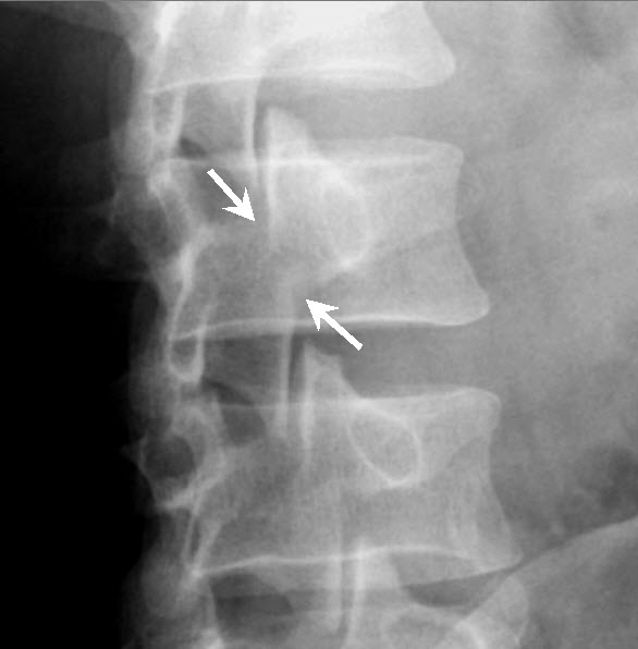
Normal anatomy-pars interarticularis. The "neck" of the "Scottie Dog" (white arrows) is intact. This is the bony bridge between the superior and inferior articulating facets of the left pars of L3.
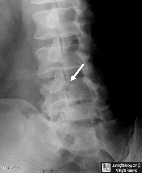
Spondylolysis. There is a break in the right pars of L4 in this patient. The "Scottie Dog" has an apparent "collar, which it should not have. The "collar" is the break in the pars.
Stinson JT. Spondylolysis and spondylolisthesis in the athlete. Clin Sports Med. Jul 1993;12(3):517-28.
Harvey CJ, Richenberg JL, Saifuddin A, Wolman RL. The radiological investigation of lumbar spondylolysis. Clin Radiol. Oct 1998;53(10):723-8.
|
|
|
