|
|
Complications of Knee Prostheses
General Considerations
- Knee prostheses typically are metallic with a polyethylene coating that forms the joint
- They may be cemented in pace or cementless
- Alignment of the components is critical for stability
- The condyles must articulate evenly with the polyethylene tibial plateau
- The tibial component is most common to loosen
- Femoral loosening more difficult to see because of overlapping metallic femoral prosthesis
- The patellar component is most prone to complications
Clinical Findings
- Painless effusion
- Retropatellar pain
- Clicking
- Instability
Imaging Findings
- Normal appearance
- Development of a thin (<2 mm) lucency at the interface of the cement and the bone or the metallic prosthesis and the bone if there is no cement
- More common around tibial component and may have a thin sclerotic line outlining them
- If, these lucencies may appear within first 6 months with cemented prosthesis or first 2 years with an uncemented prosthesis and not progress, they are normal
- Stress shielding in the distal femur
- Due to non-uniform transfer of load, a sclerotic band may appear on the lateral radiograph in the posterior aspect of the femur often associated with increased bone resorption in the non-weight bearing portion of the prosthesis

Lateral view of the knee may demonstrate minimal lucency anteriorly (white arrow)
and sclerotic band in the posterior aspect of the distal femur from possible
stress shielding (black arrow).
- Imaging of complications
- Tibial component loosens more frequently than femoral
- Signs of loosening include
- Widening of the periprosthetic lucency >2mm
- Tilting of the tibial component
- Collapse of the underlying bone
- Polyethylene beads may be shed
- Dislocation
- Particle disease is less common than in hip prostheses
- Patellar component may show wear of polyethylene component, rupture of patellar tendon
Complications
- Periprosthetic fractures may occur, especially with severe osteopenia
- Infection occurs in 0.5-2% of cases
- Most appear normal on plain radiographs
Treatment
- Revision to a long-stemmed prosthesis for loosening

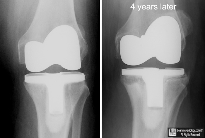
Loosening of Knee Prosthesis. Frontal radiograph of the left knee from 4 years earlier demonstrates normal post-operative appearance of prosthesis with no periprosthetic lucency (left). On right, the same knee 4 years later shows lucencies beneath the tibial portions of the prosthesis greater than 2 mm and larger than seen on the earlier study (white arrows)
For this same photo without the arrows, click here
For more information, click on the link if you see this icon 
Total Knee Arthroplasty: Postoperative Radiologic Findings. BJ Manaster. AJR 1995; 165:899-904.
|
|
|
