|
|
Hyperparathyroidism
|
|
Primary |
Secondary |
Tertiary |
Serum |
Calcium |
inc |
norm or dec |
inc |
Phosphorous |
dec |
inc |
inc |
Alkaline Phosphatase |
inc or norm |
inc or norm |
inc or norm |
Urine |
Calcium |
inc |
dec |
|
Phosphorous |
inc |
dec |
|
- Primary hyperparathyroidism
-
Overproduction of parathyroid hormone (PTH) with associated hypercalcemia
-
Most frequent cause: single adenoma in 85% of cases
-
Clinical: Bones (osteopenia, brown tumors), stones (renal calculi) and abdominal groans (ulcer disease)
-
Laboratory: increased parathormone level with hypercalcemia is diagnostic of primary hyperparathyroidism
-
Imaging of the parathyroid glands: radiolabeled technetium 99m sestamibi persists in adenomas on nuclear scans
-
Brown tumors are more common than in secondary form
-
Treatment: Symptomatic patients are usually treated by surgically removing abnormal gland(s)
- Secondary hyperparathyroidism
-
Overproduction of PTH from extrinsic stimulation most frequently from chronic renal disease
-
Most frequent cause: Parathyroid gland hyperplasia
-
Clinical: Usually the symptoms of chronic renal failure predominate
-
Laboratory: Elevated PTH with low to normal serum calcium; phosphate levels may be high in renal disease, but low in vitamin D deficiency
-
Imaging of the parathyroid glands: Imaging of parathyroid glands is not needed
-
Extra-osseous calcification is more common in secondary hyperparathyroidism than in the primary form
-
Treatment: medical management; vitamin D replacement, phosphate restriction or binding agents
- Tertiary hyperparathyroidism
-
Most frequent cause: Autonomous overproduction by all 4 parathyroid glands follows secondary hyperparathyroidism; exact cause is unknown
-
Clinical: Symptoms of hyperparathyroidism following renal transplant
-
Laboratory: Elevated PTH with hypercalcemia and frequently hyperphosphatemia
-
Imaging of the parathyroid glands: Imaging of parathyroid glands is not needed
- Treatment: Total or subtotal parathyroidectomy

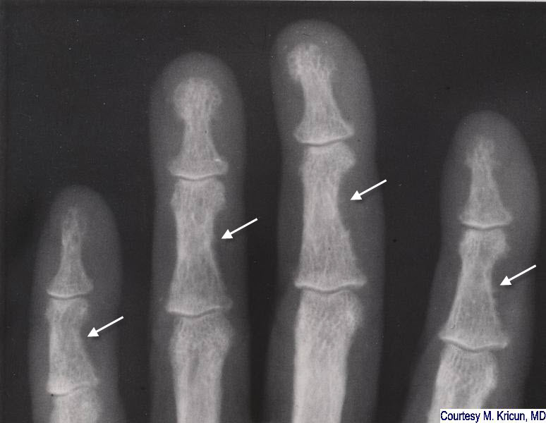
Secondary Hyperparathyroidism. White arrows point to subperiosteal resorption along the radial (lateral) aspects of the middle phalanges of the index, middle and ring fingers, a finding virtually pathognomonic for hyperparathyroidism. The cortex appears spiculated. There is acro-osteolysis (yellow arrows) of several of the terminal phalanges. A small, lytic lucency in the head of the metacarpal of the middle finger represents a brown tumor.
For more information, click on the link if you see this icon 
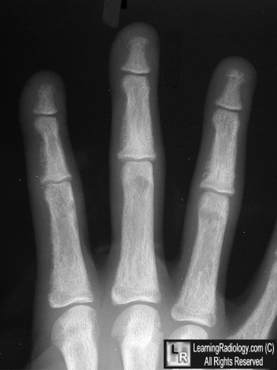
For this same photo without the annotations, click here
Secondary Hyperparathyroidism. White arrows point to subperiosteal resorption along the radial (lateral) aspects of the middle phalanges of the index, middle, ring and little fingers, a finding virtually pathognomonic for hyperparathyroidism. The cortex appears spiculated.
|
|
|
