|
|
Hemophiliac Arthritis
-
Chronic synovitis develops from repeated intra-articular hemorrhages
-
Thickened synovium produces marginal erosions
-
Multiple subchondral cysts may develop secondary to intraosseous
hemorrhage
- Conventional Imaging
-
X-ray changes due to synovial
proliferation and hyperemia
-
Widening of the intercondylar notch of the
femur
-
Chronic hyperemia produces enlargement of epiphyses
-
Secondary trabeculae are resorbed leaving linear striations in the bone
-
Sometimes hemosiderin in soft tissues may make them appear dense
-
From the increased blood to the epiphyses,
the epiphyses may appear too early, grow too large, and fuse early
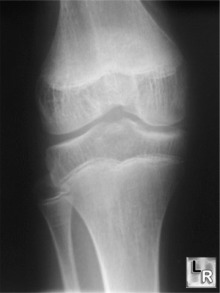
Hemophiliac Arthritis. There is widening of the intercondylar notch,
accentuation of the
trabeculae and enlargement of the medial epicondyle
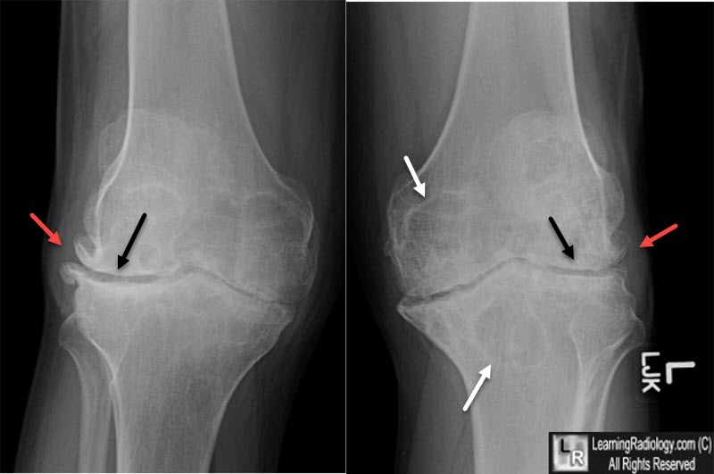
Hemophiliac Arthritis. There are changes of osteoarthritis of both knees in a 37 year-old. The age of the patient should raise the diagnosis of secondary osteoarthritis. The patient was known to have hemophilia. There are marginal osteophytes (red arrows), narrowing of the joint spaces with subchondral sclerosis (black arrows) and large subchondral cysts (white arrows).
|
|
|
