|
|
Soft Tissue Hemangioma
General Considerations
- Common, benign, soft-tissue tumors (7% of all benign soft tissue tumors)
- Women more commonly affected than men
- Most commonly diagnosed soft tissue tumor in children
- They can arise from skeletal muscle, skin, subcutaneous tissue and synovial tissue
- Histologic varieties include capillary, cavernous, arteriovenous, venous and mixed
- Capillary hemangiomas are most common, usually found in childhood
- Most spontaneously disappear
- Cavernous hemangiomas are larger and occur in later life
- Occur frequently in muscle
- Calcification is common
- Arteriovenous hemangiomas can cause shunting between the arterial and venous system
- Venous hemangiomas are found deep, such as in the retroperitoneum and mesentery
Clinical Findings
- Smooth, palpable mass
- Can increase in size during pregnancy
- Chronic pain in 60%, may be worsened with exercise
- Bluish discoloration of skin
Imaging Findings
- Radiographs may be normal or may show a soft tissue mass containing multiple phleboliths (especially in cavernous hemangiomas)
- The mass may produce periosteal reaction or cortical thickening in adjacent bone
- On unenhanced CT, smaller phleboliths may be visible
- Enhanced CT will show numerous, serpentine vessels, prominently enhancing
- There may be an increase in adjacent fat
- Doppler US may show low resistance arterial flow
- MR is the current standard for imaging
- All sequences show a heterogeneous mass
- T1-weighted images show increase in fat around circumference of lesion
- On T2-weighted images, the central portion of the mass shows high signal intensity
Treatment
- Asymptomatic hemangiomas usually are not treated
- Symptomatic lesions may be removed surgically or by laser
- Surgery is frequently preceded by embolization to reduce bleeding
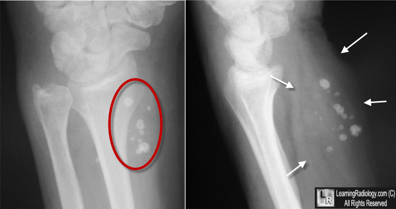
Hemangioma, soft tissue. There is a soft tissue mass (white arrows) which contains numerous spherical calcifications characteristic of phleboliths (red oval). The combination is characteristic of a hemangioma of the soft tissue, most likely of the cavernous variety.
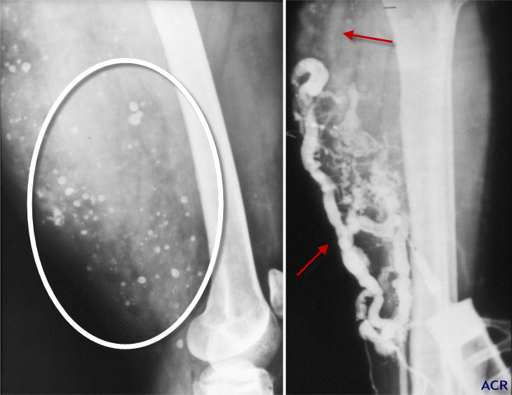
Hemangioma, soft tissue. There is a huge soft tissue mass containing innumerable phleboliths in the soft tissue of the thigh (white oval) that is seen to contain numerous enlarged and disordered arterial and venous channels (red arrows) on the angiogram done on the right.
Soft-Tissue Cavernous Hemangioma. Kristina I. Olsen, G. Scott Stacy, and Anthony Montag. RadioGraphics, May 2004, Vol. 24, Issue 3.
|
|
|
