|
|
Bone Infarct
Medullary Bone Infarct, Osteonecrosis
General Considerations
- Usually found within the intramedullary cavity of the metaphysis or diaphysis of bone
- When osteonecrosis occurs in the epiphysis, it is called avascular necrosis
- In long bones, their calcification is similar to an intramedullary bone infarct
- Bone infarcts tend to have a well-circumscribed, sclerotic margin
- Due to interruption of the blood supply from numerous causes including:
- Sickle cell disease
- Polycythemia
- Arteritis, as in connective tissue diseases
- Trauma
- Idiopathic
- Excessive exogenous or endogenous steroids
- Alcoholism and pancreatitis
Imaging Findings
- Intramedullary calcification will be seen on conventional radiographs after months or years
- Frequently has a well-defined “membrane” or “shell” surrounding it (unlike enchondromas)
- There may be associated periostitis
- MRI is much more sensitive to ischemic changes and may show findings in 6-12 hours
- On MRI, the signal in the center of a chronic bone infarct is similar to normal marrow; in acute infraction, the T1 signal is decreased
- “Double line sign” on T2-weighted MRI is due to hyperintense central ring of granulation tissue and hypointense peripheral ring that is sclerotic
Differential Diagnosis
Prognosis
- Very rarely may de-differentiate into a malignancy such as osteosarcoma or fibrosarcoma
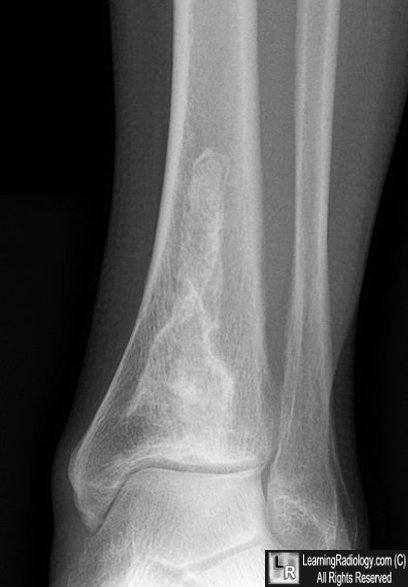
Chronic Bone Infarct. There is a well-defined, intramedullary calcification in the meta-diaphysis of the distal tibia with a "shell-like" outer calcified membrane characteristic of an old bone infarct.
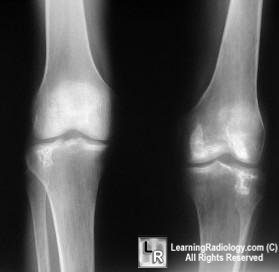
Densities in both proximal tibias an distal femurs represent areas of avascular necrosis in a patient with sickle cell disease.
|
|
|
