|
|
"Shin Splints"
General Considerations
- Catch-all term used to describe almost any pain in tibia
- The most common cause of shin splints is medial tibial stress syndrome
- Seen in military personnel and amongst athletes
- Exercise induced pain and tenderness along posterior-medial border of tibia
- Usually seen in athletes who increase their training
Clinical Findings
- Dull, aching pain on posterior and medial aspect of calf
- Frequently occurs at beginning of a sports season
Imaging Findings
- Conventional radiographs are normal
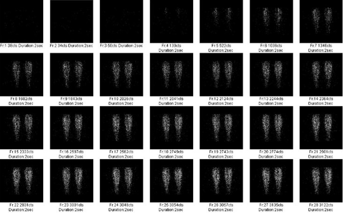
- Nuclear medicine bone scans
- Blood pool images are normal
- Long, longitudinally-oriented uptake on delayed images in posterior tibia
- Tracer uptake varied along length of lesion
- Must obtain both lateral and medial views
- Findings differentiate shin splints from stress fractures
Differential Diagnosis
- Stress fracture
- Chronic compartment syndrome
- DVT
- Muscle strain
Treatment
- Rest
- Ice
- Compression
- Elevation
 
"Shin Splints." White arrows point to linear region of increased uptake in tibia on delayed radionuclide"Triple phase" bone scan images consistent with so-called shin splints in this young athlete. The blood
flow study shows equal flow in both calves (red circle)
.
For more information, click on the link if you see this icon 
For these same photos without the annotations, click here and here
The Specific Scintigraphic Pattern of Shin Splints in the Lower Leg: Concise Communication. Lawrence E. Holderand and Roger H. Michael. J Nuci Med 25: 865-869, 1984
|
|
|
