|
|
Lacunar Skull
Luckenschadel Skull
General Considerations
- Bone dysplasia of skull consisting of multiple oval lucencies separated by dense, bony ridges
- Associated with
- Neural tube defects, especially myelomeningocele
- Chiari II malformation
- Encephalocele
- Not related to degree of concomitant hydrocephalus
- Inner table more affected than outer
Clinical Findings
- Present at birth
- Unrelated to increased intracranial pressure
Imaging Findings
- Well-defined lucent areas in calvarium representing nonossified fibrous bone
- Lacunae are bounded by normally ossified bone
- Most prominent in parietal bones
- Small posterior fossa associated with Chiari II malformation
Differential Diagnosis
- Normal convolutional markings seen during a period of rapid brain growth (3 to 7 years).
- Increased convolutional markings associated with synostosis at an older age
- Increased intracranial pressure - beaten-silver appearance; closed sutures; may have abnormal skull size
Treatment
- Appearance resolves spontaneously by age 6 months
Prognosis
- Spontaneously disappears by 4-6 months old

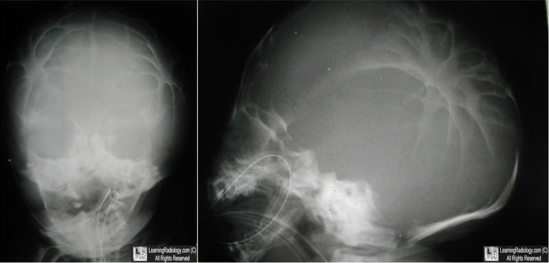
Lacunar Skull. There are multiple focal areas of radiolucency in the skull (white arrows)
bounded by more normal, dense bony ridges. The child had a known myelomeningocele.
For this same photo without the arrows, click here
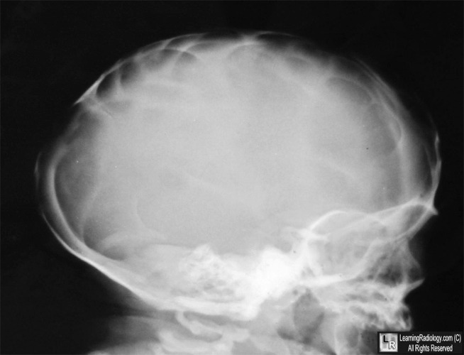
Luckenschadel. This newborn skull is markedly dysplastic with little in the way of normal, dense bony ridges.
For more information, click on the link if you see this icon 
The Infant Skull: A Vault of Information. RB J Glass, S K Fernbach, KI Norton, PS Choi and TP Naidich. March 2004 RadioGraphics, 24, 507-522.
|
|
|
