|
|
Ureterocele
Ureterocoele
General Considerations
- Congenital saccular outpouching of distal ureter into the urinary bladder
- Most common in white females
- Most are left-sided but about 10% are bilateral
- Most ureteroceles found in adults are orthotopic whereas those in children are far more commonly ectopic
- In a duplex system, 80% of ureteroceles are associated with the upper pole moiety
- Upper pole frequently obstructs
- Lower pole undergoes vesico-ureteral reflux
Duplex collecting Systems |
Upper pole more prone to obstruction |
Inserts medially and inferiorly to normal ureter |
Associated with ureterocele |
Lower pole ureter is associated with reflux |
- Most ureteroceles are associated with complete duplication of the ureter
- Believed due to obstruction of ureteral orifice during embryogenesis
Clinical Findings
- In both children and adults they are often found incidentally
- Symptoms are related to the obstructive nature of ureteroceles or to their ectopic insertion
- Hydronephrotic kidney may produce and abdominal mass
- Symptoms include
- Recurrent cystitis
- Bladder outlet obstruction
- Failure to thrive
- Hematuria
- Calculus formation
- Abdominal pain
Type |
Description |
Single system |
Single kidney; single collecting system; single ureter |
Duplex-system |
Duplicated collecting system; two ureters |
Orthotopic (intravesical) |
Orifice of ureterocele is located in normal anatomic position in bladder |
Ectopic (extravesical) |
Orifice is located in bladder neck, urethra |
Imaging Findings
- Stones in a ureterocele occur in adults
- CT urogram
- Has primarily replaced the conventional intravenous pyelogram
- Duplicated collecting system
- Hydroureteronephrosis
- If upper pole is obstructed, it may not opacify
- Upper pole moiety produces drooping lily sign by displacing lower pole downward and laterally
- Ureter from upper pole moiety inserts in a more inferior and medial location than lower pole moiety (Weigert-Meyer Rule)
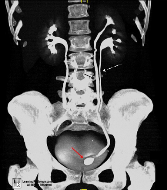
- Cobra-head appearance of the dilated distal ureter
- Elliptical collection of contrast in ureterocele surround by this radiolucency of wall of ureterocele
- Opaque calculi on the ureterocele will be seen on the pre-contrast scout image
- Non-opaque calculi in ureteroceles will be seen on the post-contrast studies as filling defects within the ureterocele
- Ultrasound (US)
- Study of choice in both adult and pediatric population
- Fluid filled cystic mass within the bladder
- Hydroureteronephrosis
- Magnetic Resonance urography an excellent tool to evaluate poorly functioning moiety when associated with ectopia
Differential Diagnosis
- Bladder diverticula
- Mesonephric duct cysts
- Pseudoureteroceles
- Edema surrounds distal ureter from inflammation from stone, radiation cystitis or possible transitional cell ca of the bladder
Treatment
- Small, asymptomatic ureteroceles causing no obstruction may require no treatment
- Surgery may be indicated if any of the following are present
- Recurrent urinary tract infections
- Ureteral calculi
- Intractable pain
- Compromise of renal function
- Surgery includes
- Transurethral unroofing of the ureterocele
- Upper pole heminephrectomy and partial ureterectomy
- Excision of ureterocele with reimplantation of ureter
Complications
- Vesico-ureteral reflux following surgical correction
- Incontinence from damage to bladder neck
- Injury to contralateral ureter
- Renal hypertension

Ureterocele. A reformatted coronal image from a CT urogram demonstrates a partially duplicated collecting system on the left side with two ureters (black and white arrows) that join at the pelvic inlet and insert as one ureter into the bladder. At the insertion of the ureter, there is a saccular collection of contrast with a halo of lucency surrounding it characteristic of a ureterocele.
For this same image without the annotations, click here
Ureterocele eMedicine Minevich, E; Tackett, L; Choe, J
|
|
|