|
|
Retrocaval Ureter
Circumcaval Ureter
- Also known as “circumcaval ureter”
- Abnormality in embryogenesis of IVC
- Results from abnormal persistence
of right subcardinal vein positioned ventral to ureter
in the definitive IVC
- Developing right ureter courses
behind and medial to the IVC
- Incidence
- 0.07%
- Male to female ratio of 3:1
- Clinical findings
- Symptoms of right ureteral
obstruction
- Imaging findings
- Normal course of ureters
- About the width of your thumb
lateral to the lumbar vertebral pedicles
- About the width of two fingers
medial to pelvic brim in true pelvis
- With retrocaval ureter
- Right ureter’s course swings
medially over pedicle of L3/4
- Then exits anteriorly between
IVC and aorta returning to its normal position
- Produces varying degrees of
proximal hydroureteronephrosis
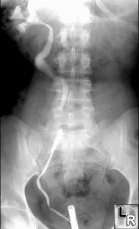
Retrograde pyelogram of right ureter
demonstrates displacement of the ureter which passes
medial to the pedicle ft the level of L4. The ureter is
slightly dilated proximal to this point and returns to a
normal position distal to its retrocaval placement.
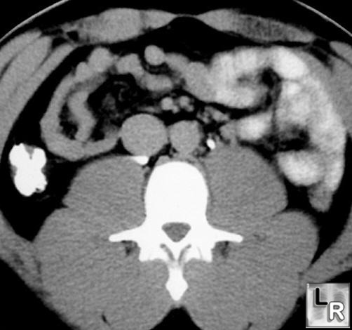
CT scan below the level of the kidneys
demonstrates a more medial
retrocaval placement of the right ureter.
- Retrocaval ureter can be associated
with Turner’s syndrome
Causes of Ureteral
Deviation or Displacement |
Medial Displacement or Deviation |
Upper ureter |
Lower ureter |
Retrocaval ureter |
Lymphadenopathy |
Retroperitoneal fibrosis |
Iliac artery aneurysm |
|
Bladder diverticulum |
|
Post-surgical (esp. AP resection) |
|
Pelvic lipomatosis |
Lateral Displacement or Deviation |
Upper ureter |
Lower ureter |
Lymphadenopathy |
Pelvic mass, e.g. uterine fibroids |
Aortic aneurysm |
|
Retroperitoneal hematoma |
|
Herman, T and McAlister, W: Radiologic Clinics of North
America. Vol. 29:2, March, 1991
|
|
|
