|
|
Congenital Megacalyces
General Considerations
- Rare; more common in males
- Congenital
- Usually unilateral
- Underdevelopment of medullary pyramids
- No obstruction
- The renal pelvis and ureter are usually normal
- Rarely associated with megaureter which is an obstructive uropathy occurring near the ureterovesical junction
Clinical Findings
- Near-normal renal function
- Often symptom-free
- Stasis in calyces may predispose to infection and stones
Imaging Findings
- Since the medullary pyramids are underdeveloped, the calyx is enlarged and flattened or convex instead of concave
- Kidney may be larger than normal
- Frequently more calyces than normal
- Calyces are facetted, fitting together as pieces of a puzzle
- Renal medulla is thinner than normal but cortex retains its normal thickness and function
Differential Diagnosis
- Hydronephrosis – the renal pelvis is general not enlarged with megacalyces but is with hydronephrosis
Treatment
- Not progressive
- No treatment is usually necessary
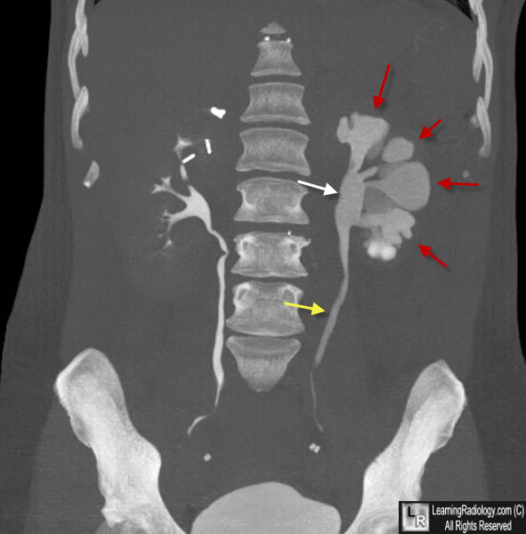
Congenital Megacalyces. Maximum Intensity projection Image of a CT urogram shows numerous dilated, polygonal-shaped calyces in the left kidney with convex outer margins (red arrows). The calyces fit together like pieces of a puzzle. The renal pelvis (white arrow) is not dilated and the ureter is normal in size (yellow arrow) helping to differentiate this from a cause of obstructive hydronephrosis.
The Coexistence of Congenital Megacalyces and Primary Megaureter. B Vargas and RL Lebowitz. AJR 147:313-316 August 1986.
Talner LB, Gittes RF. Megacalyces. Clin Radiol. 1972;23(3):355-361.
|
|
|
