|
|
Horseshoe Kidney
General Considerations
- Most common fusion abnormality of the kidney
- Fusion is at lower pole in 90% of cases
- The majority of each kidney lies on its own side of the spine
- In crossed fused ectopia, both fused kidneys lie on the same side of the spine
- May occur as isolated anomaly or with other anomalies (1/3), such as
- Reflux, hypospadias, retrocaval ureter, imperforate anus, Meckel diverticulum
Clinical Findings
- About 1/3 are asymptomatic
- When present, symptoms stem from hydronephrosis, stones, infection
- Vague abdominal pain may be most common presenting symptom
Imaging Findings
- CT is probably study of choice now
- Isthmus (fused portion) is usually functioning renal parenchyma but can be fibrous
- Isthmus is anterior to aorta and inferior vena cava and posterior to inferior mesenteric artery
- Renal scintigraphic scanning may also demonstrate whether the isthmus contains functioning tissue of not
- Sometimes incidentally diagnosed on bone scans
- Renal pelvis is usually rotated anteriorly and ureters arise anteriorly or laterally
- If both upper and lower poles fuse, called pancake or doughnut kidney
- In only 1/3 of cases is there a single renal artery for each kidney
- In most cases, one or both kidneys have 2-3 renal arteries and isthmus receives its supply from renal artery or aorta
- Axis of kidneys is tilted such that upper poles are more lateral than lower poles
- Lower pole calyces lie medial to the ureters
- Renal pelvis may be extrarenal and large
- There may be evidence of hydronephrosis from UPJ obstruction
Treatment
- UPJ obstruction may be treated surgically
- Stones may be treated with lithotripsy
Complications
- Ureteropelvic junction obstruction
- Recurrent infections (frequent)
- Recurrent calculus formation (frequent)
- Increased incidence of Wilms tumors, transitional cell carcinoma and renal carcinoids
Prognosis
- Normal survival rate with isolated abnormality
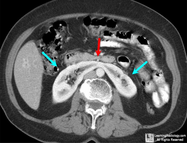
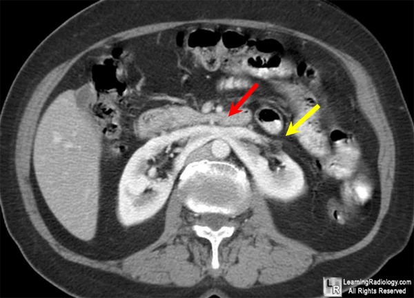
Horseshoe Kidney. Enhanced axial CT scan of abdomen at two near-contiguous levels
shows
a horseshoe-shaped kidney (blue arrows) which has functioning
renal parenchyma in the isthmus that
crosses the midline (red arrows).
The ureter is seen anteriorly on the left (yellow arrow).
For more information, click on the link if you see this icon 
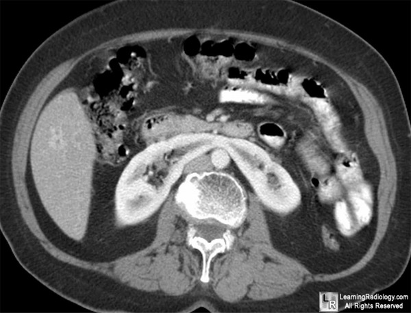
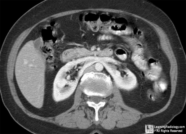
For these same photos without the arrows, click here and here
eMedicine Horseshoe Kidney Abid Irshad, A; Ackerman, S; Ravenel, J
|
|
|
