|
|
Target Sign
Bowel Inflammation
General Considerations
- Three concentric circles formed by the layers of the bowel wall in inflammatory disease of the bowel and seen on contrast-enhanced CT
- The inner and outer rings of high attenuation are presumed to represent hyperemia of the mucosa and muscularis propia and/or serosa, respectively
- The middle low attenuation ruing is presumed to represent submucosal edema which produces thickening of the bowel wall
- Best seen during late arterial, early venous phase
- Originally described with Crohn disease, it can be seen in any inflammatory disease of the bowel, including
- C. Difficile colitis
- Ischemic bowel disease
- Radiation-induced colitis
Clinical Findings
- Depend on underlying cause
Imaging Findings
- Most common CT finding in inflammation of the bowel is bowel wall thickening
- Intensity of enhancement correlates with severity of disease
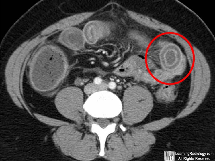
Target Sign. Contrast-enhanced axial CT of lower abdomen in patient with Crohn disease demonstrates three concentric circles (red circle) representing hyperemic mucosa and muscularis/serosa as inner and outer high density rings and submucosal edema in the central, low density ring.
For more information, click on the link if you see this icon 
For this same photo without the annotations, click here
The Target Sign: Bowel Wall. Ahualli, J. February 2005 Radiology, 234, 549-550.
|
|
|
