|
|
Liver Laceration
- Most frequently injured abdominal organ after spleen
- Most often due to deceleration injuries
- Often seen in association with
- Right-sided rib fractures
- Right-sided pneumothorax
- Right lung contusion
- Injuries to the right kidney or adrenal gland
- Injuries include
- Subcapsular hematoma
- Laceration
- Intrahepatic hematoma
- Contusion
- Right lobe more often injured than left
- Injury to left lobe associated with injury to duodenum, pancreas, transverse colon
- More often due to direct blows to the epigastrium
- High association with injuries to other organs
- 45% with liver injuries have splenic injury
- Subcapsular hematomas
- Lenticular configuration
- Flattens adjacent liver
- Often adjacent to rib fracture
- Most occur in antero-lateral aspect of right lobe
- Liver Laceration
- Non-enhancing region, linear or branching
- Frequently parallel hepatic vein
- Hypodense wedge extending to liver surface
- Focal hepatic devascularization
- Periportal tracking of blood
- Frequent finding
- Sometimes only evidence of injury
- Due to dissecting hemorrhage
- Bile
- Dilated periportal lymphatics
- Hematoma
- Higher attenuation than surrounding liver on unenhanced CT scan
- Lower attenuation than surrounding liver on enhanced CT scan
- Central high attenuation region containing clot
- Hepatic vein laceration usually involves right hepatic vein near vena cava
- Contusion
- Rare lesions
- Low attenuation area compared to normally enhanced liver
- Do not disrupt major portal or hepatic venous structures
- Hemoperitoneum
- Complications
- Delayed rupture (rare)
- Hemobilia
- Arteriovenous fistula
- Pseudoaneurysm
- Biloma
- Superinfection of hematoma
- Pitfalls
- Adjacent rib artifacts-beam-hardening
- Mimics laceration
- Adjacent to ribs
- Fade as they become farther from rib
- Linear artifact from air-contrast level in stomach
- Fatty liver with laceration or hematoma can be missed
- Clue- look at intrahepatic ducts and vessels
- Treatment
- Conservative treatment in up to 80% in adults and almost all children
- Monitor hemodynamic state of patient
- Transcatheter embolization possible for bleeders
- Healing
- Contusions may clear in 5-7 days
- Subcapsular hematomas may increase in size initially before clearing
- Lacerations can heal within weeks but small, residual bilomas are common
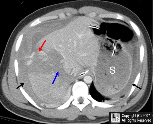
Liver laceration with extravasation. An enhanced axial CT scan of the upper abdomen shows a large laceration through the right lobe of the liver (blue arrow), blood in the peritoneal cavity (black arrows) and active extravasation of the intravenous contrast (red arrow). The stomach is labeled "S."
For additional information about this disease, click on this icon if above.
For this same photo without the arrows, click here
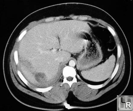
Liver Laceration. Contrast-enhanced CT of abdomen shows linear
low-attenuation defect
crossing the posterior aspect of the right lobe
of the liver representing a
laceration.
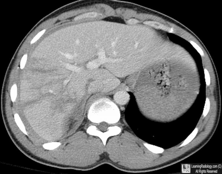
Liver Laceration. Contrast-enhanced CT of abdomen shows multiple linear
low-attenuation defects
crossing the right lobe
of the liver representing
lacerations.
|
|
|
