|
|
Gastroschisis
General Considerations
- Herniation of abdominal contents through a small (2-5 cm) defect in the abdominal wall, usually just to the right of the umbilicus
- Usually involves small bowel
- Herniation of other visceral organs is rare
- No surrounding membrane
- There are usually no associated congenital defects
Clinical Findings
- Maternal serum alpha-fetoprotein (AFP) and amniotic fluid AFP levels are elevated
- There may be polyhydramnios
Imaging Findings
- Antenatal ultrasound is the study of choice
- Usually found in the second trimester using antenatal sonography with loops of bowel floating in the amniotic fluid
- Normal insertion of the umbilical vessels into the fetal abdomen
Differential Diagnosis
- Omphaloceles are herniations into the base of the umbilical cord surrounded by a membrane
- They are more common and are associated with other congenital abnormalities
- The liver is exteriorized
- Physiologic bowel herniation occurs at 10-13 weeks
Treatment
- Surgical repair is usually performed within the first day after delivery to avoid infection
Complications
- Bowel obstruction
- Bowel perforation
- Peritonitis
- Fetal growth restriction, 50% of fetuses being small for their gestational dates
Prognosis
- Mortality rate is approximately 17%
- Surgical repair is usually performed within the first day after delivery to avoid infection

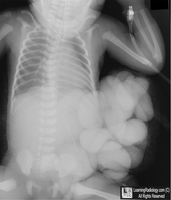
Gastroschisis. Newborn whose mother had not undergone antenatal testing
shows multiple loops of small bowel (white arrow) outside of the abdomen
having herniated through the abdominal wall. The orogastric tube tip (black arrow) extends farther inferior than normal.
For this same photo without the arrows, click here
For more information, click on the link if you see this icon 
|
|
|
