|
Colonic Polyps
•
The majority of colorectal polyps are inflammatory or metaplastic and usually
5mm or less in diameter and have no malignant potential; the majority of larger lesions are adenomatous polyps
•
Predisposing conditions:
• Prior detection of colonic polyp
• Cancer of the colon
• Family history of polyps or colon cancer
• Inflammatory bowel disease (ulcerative colitis, Crohn’s)
• Familial polyposis
• Peutz-Jeghers’s Syndrome
• Gardener’s syndrome
•
Incidence rises with age
• About 3% in 3rd decade
• 10% in 7th decade
• 26% in 9th decade
• About 11% overall in all ages
•
Most (60%) occur in rectum or sigmoid
•
Types
Type
|
Incidence
|
Description
|
Malignant
Potential
|
Tubular
adenoma
|
75%
|
Cylindrical
glandular structure lined by columnar epithelium
|
<10mm=1%;
1-2cm=10%;
>2cm=35%
Least
malignant
|
Tubulovillous
adenoma
|
15%
|
Mixture
between tubular and villous adenoma
|
<1cm=4%;
1-2cm=7%;
>2cm=46%
|
Villous
adenoma
|
10%
|
Infolding
of papillary projections of glands
|
<1cm=10%;
1-2cm=10%;
>2cm=53%
Most
malignant
|
•
Overall size versus malignancy: <1cm=1%; 1-2 cm=10%; >2cm= 46%
•
Symptoms
• Most are asymptomatic
• Some may have diarrhea, especially villous tumors in rectum
• May also produce hypokalemia
• Abdominal pain (may be 2° to intussusception in a few)
• Rectal bleeding correlates with size and may be seen in as many as
67%
•
Imaging
• Rate of detection of polyps less than 1 cm is higher with air
contrast
• Rate of detection of polyps 1cm or greater is about equal with
single vs. double contrast
• From 1/4 to 1/2 of patients with one polyp have a synchronous
lesion
• May be sessile or on a stalk
• Bowler hat sign (sessile
polyp viewed in profile on A/C exam)
• Target sign (polyp with
stalk viewed en face)
• Stalk >2cm is almost always benign
•
Differentiating Benign from Malignant polyps
• Size (see above)
• Presence of a stalk
• Lesions on a stalk have less of a chance of being malignant than a
sessile lesion of the exact same size
• Even when malignant polyps have a stalk, the chance of spread to
regional nodes is low
• Surface contour
• Not really reliable
• An ulceration is more consistent with ca
• Dimpling at the base suggests ca
•
If greater than 3cm in size, barium trapped within interstices suggests
villous tumor
• Location
• Does not help
• Growth
• Any polyp which undergoes an interval increase in size should be
removed
Juvenile
Polyps
•
Classified as cystic hamartomas by some and inflammatory retention cysts by
others
•
No malignant potential
•
Most occur as isolated colonic lesions in children less than 10 years
•
Most are solitary
•
Rectal bleeding is the most common symptom
•
Most occur in the rectum or sigmoid
•
Since they have a tendency to autoamputation, they are usually not removed
Hyperplastic
Polyps
•
No malignant potential
•
Mucous glands lined by a single layer of columnar epithelium
•
Usually located in rectum
•
Usually less than 5mm in diameter

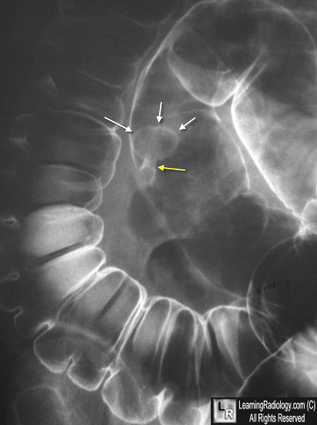
Colonic Polyp. This is a close-up of the sigmoid colon from an air-contrast (double-contrast) barium enema. Outlined by a thin layer of barium is a large pedunculated polyp. The yellow arrow points to its stalk and the white arrows to the head of the polyp.
|