|
|
Imaging Findings in Chronic Pancreatitis
General Considerations
- Imaging plays a key in the diagnosis and management of chronic pancreatitis.
- Uncomplicated chronic pancreatitis is usually treated symptomatically.
- Complications such as pseudocysts, abscess, and malignancy may require minimally invasive therapy or surgery.
- Acute pancreatitis and chronic pancreatitis are generally assumed to be different disease processes, with most instances of acute pancreatitis not leading to chronic disease.
- Chronic calcifying pancreatitis is almost always related to alcoholism.
- Chronic obstructive pancreatitis is usually more focal than those in the other forms, and in most patients, the changes involve only the portion of the pancreas in which ductal drainage is impaired.
- Calcification is unusual.
- The pancreatic duct is invariably dilated.
Conventional Radiography
- May show punctate or coarse calcifications in up to 60% of those with the disease and are pathognomonic for alcoholic chronic pancreatitis
- They primarily represent intraductal calculi, either in the main pancreatic duct or in the smaller pancreatic ducts.
- The calcification may have a focal, segmental, or diffuse distribution
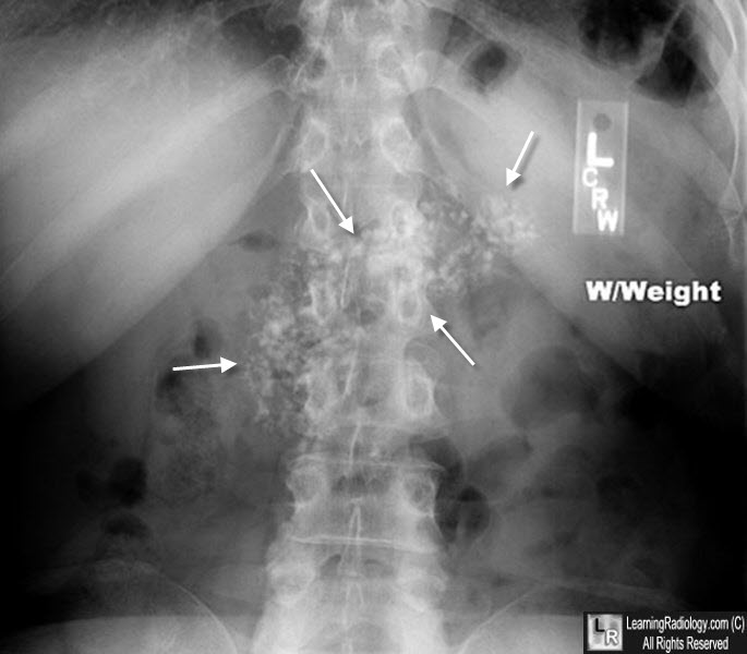
Chronic Pancreatitis. There are innumerable calcifications in the region of the pancreas. Their amorphous appearance speaks to them forming in a sold organ.
CT
- Diameter of the main pancreatic duct is greater than 5 mm in the head and 2 mm in the tail
- Most sensitive for demonstrating pancreatic calcifications
- Focal enlargement or atrophy of the pancreatic parenchyma
- Obliteration of the peripancreatic fat usually indicates acute exacerbation
- Main pancreatic duct may show a chain-of-lakes appearance due to alternating stenoses and dilatation
- Obstruction of the common bile duct with gradual tapering
- Pancreatic pseudocysts
- In approximately 50% of patients with chronic calcific pancreatitis, the pancreatic parenchyma contains cysts of varying sizes
- Ascites
- Splenomegaly
- Obstruction of the GI tract
US
- Irregular contour of the pancreas seen in 45-60%
- Diffuse enlargement occurs in 27-45%
- Hypoechoic early in disease
- Later is disease the pancreas may become heterogeneous, with areas of increased echogenicity along with focal or diffuse enlargement
- Focal enlargement is detected in 12-32%
- Peripancreatic fascial thickening and blurring of the pancreatic margins is seen in approximately 15% of patients
- Pseudocysts may occur
- Calcification in the gland results in densely echogenic foci, which may shadow.
- The pancreatic and common bile ducts may be dilated and the pancreatic duct may be beaded in appearance
- In the late stages of the disease, the pancreas becomes atrophic and shrinks.
- Small, echogenic pancreas with a heterogeneous echotexture
- Pancreatic duct remains dilated
MRI
- Magnetic Resonance Cholangiopancreatography (MRCP) may depict the beaded appearance of the pancreatic duct
- Pancreatic duct calculi are depicted as round filling defects
- Fat-suppressed T1-weighted images usually show a loss of signal intensity
MedScape. Chronic Pancreatitis Imaging. AN Khan and AJ Sheen
|
|
|
