Precancerous
states
•Pernicious
anemia—18X normal population
•Adenomas of the stomach—especially those over 2cm
•Atrophic Gastritis—disputable
•Hiatal hernia—disputable
•Gastric stumps for ulcer disease (Bilroth II>Bilroth I)
•Achlorhydria
Histology
• Adenocarcinoma (95%)
• Rarely, squamous cell
Morphology
• Polypoid/fungating carcinoma
• Ulcerating/penetrating carcinoma (70%)
• Infiltrating/scirrhous type=linitis plastica
• Superficial spreading type-confined to mucosa/submucosa-NOT linitis
plastica
Metastases
• Along peritoneal ligaments
∆ Gastrocolic ligament to transverse colon
∆ Gastrohepatic and hepatoduodenal to liver
• To lymph nodes
∆ Locally
∆ Lymphangitic to lungs
• Hematogenous
∆ Liver (most common)/adrenals/ovaries/bones
• Peritoneal seeding
∆ Rectal wall=Blumer shelf
• Left supraclavicular node=Virchow’s node
•
Overall 5 year survival 5-18%
Malignant
ulcer—is
a carcinoma which presents with the radiographic appearance of an ulcer niche;
these have the radiographic appearance of a benign ulcer but demonstrate
microscopic foci of malignancy, usually at the edge of the ulcer
Ulcerating
malignancy—is
a carcinoma having sufficient bulk to present as a mass which also contains a
persistent collection representing an ulcer; the mucosa is frequently nodular
and the folds do not radiate to the base of the ulcer
Linitis
plastica (scirrhous carcinoma)—is
a diffuse involvement of the wall of the stomach, frequently with flattening
of the mucosa, and poor distensibility and contraction of the wall; usually
associated with significant fibrosis and muscular hypertrophy; very frequently
a signet ring cell type

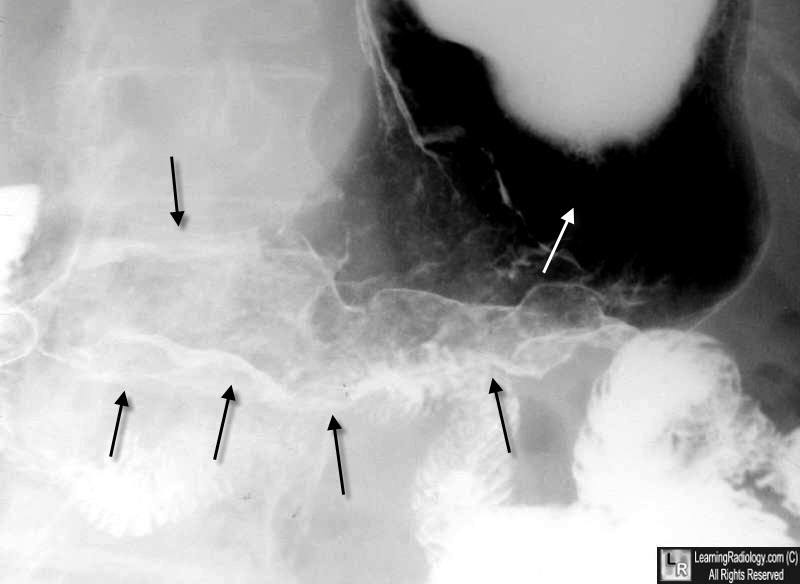
Carcinoma of the Stomach, UGI. A double-contrast upper GI of the stomach shows a markedly rigid and non-distensible body and antrum of the stomach (black arrows) which did not change on multiple views. The proximal portion of the stomach (white arrow) does distend suggesting the tumor involves only the distal stomach..

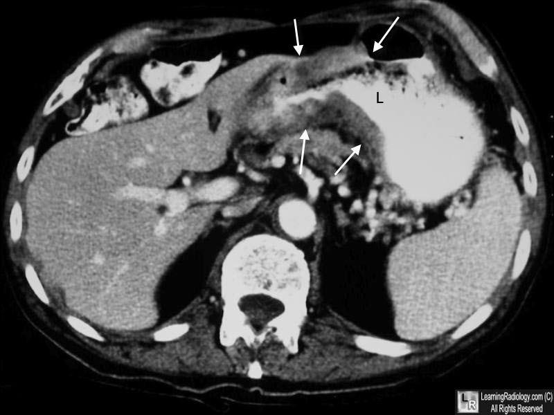
Carcinoma of the Stomach, CT. The gastric wall in the region of the antrum of the stomach shows marked soft-tissue thickening (white arrows) while the lumen (L) is constricted by the surrounding mass.
|