|
|
Intracranial Tuberous Sclerosis
General Considerations
- Rare, multi-system genetic disease
- Classical triad consists of mental retardation, seizures and adenoma sebaceum
- Cutaneous hamartoma (angiofibroma) is known as adenoma sebaceum
- Hamartomas in the brain (tubers)
- Other hamartomatous lesions may affect the heart, lungs, kidneys (including angiomyolipomas), gastrointestinal polyps and bones
Clinical Findings
- Neurologic findings may include seizures, autism and developmental delays
- Blindness
- Hemiparesis
Imaging Findings
- Cortical tubers are developmental abnormalities that occur in the cerebral cortex, mainly in the frontal lobe (50%)
- Calcify frequently after the age of two
- On MRI, high on T2 and low on T1 with infrequent enhancement
- Subependymal nodules usually are multiple
- Most are eventually associated with calcification (88%)
- On MRI, high on T1 and isodense to high on T2
- Variable enhancement
- Subependymal giant cell astrocytomas (1-26%)
- Occur between 8-18 years of age at peak
- Usually large and occur mostly at Foramen of Monro leading to hydrocephalus
- Pathologically and biologically, they behave more like a benign lesion
- On MRI, enhance aggressively
- Cystic or radial band-like lesions in white matter (40-90%)
- White matter abnormalities are related to almost all cortical tubers
- Radial Bands- linear bands on MRI which radiate from the periventricular white matter to the subcortical region
- Specific for tuberous sclerosis
- Chiari malformations
- Cerebellar atrophy
- Aneurysms
- Arachnoid cysts
- Infarcts
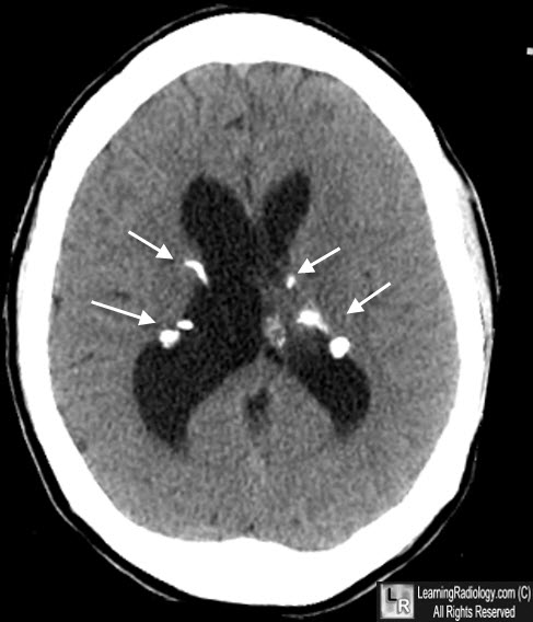
Tuberous Sclerosis. Unenhanced CT shows multiple, calcified nodules in a periventricular, subependymal distribution bilaterally in dilated lateral ventricles, characteristic of tuberous sclerosis.
Pictorial Review of Tuberous Sclerosis in Various Organs. Shigeaki Umeoka, Takashi Koyama, Yukio Miki, Mikio Akai, Kazushige Tsutsui, and Kaori Togashi. Radiographics, November-December 2008, Volume 28, Issue 7
|
|
|
