|
|
Hemorrhagic Stroke
- Hemorrhage occurs in about 15% of strokes
- Hemorrhage is associated with a higher morbidity and mortality than ischemic stroke
- In the majority of cases, there is associated hypertension
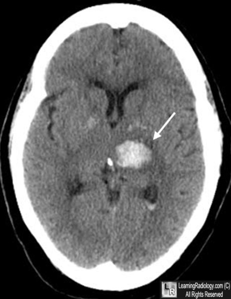
- About 60% of hypertensive hemorrhages occur in the basal ganglia
- Other areas involved are the thalamus, pons and cerebellum
- The decision to utilize thrombolytic therapy is based on algorithms formulated by the initial non-enhanced CT scan findings
- Treatment should be initiated within one hour after the patient arrives at the hospital to provide its maximum benefit
- Recognizing intracerebral hemorrhage (in general)
- Freshly extravasated whole blood with a normal hematocrit will be visible as increased density (hyperdensity, hyperattenuation) on non-enhanced CT scans of the brain
- This is due to the protein in the blood (mostly hemoglobin)
- Dissection of blood into the ventricular system can occur in hypertensive intracerebral bleeds
- As the clot begins to form, the blood becomes denser for about 3 days because of dehydration of the clot
- After the 3rd day, the clot decreases in density and becomes invisible over the next several weeks
- The clot loses density from the outside in so that it appears to shrink
- After about two months, only a small hypodensity may remain

Intracerebral hemorrhage, acute. Freshly extravasated whole blood, as this bleed into the thalamus (thin white arrow) will be visible as increased density on non-enhanced CT scans of the brain due primarily to the protein in the blood (mostly hemoglobin). As the clot begins to form, the blood becomes denser for about 3 days because of dehydration of the clot. After the 3rd day, the clot gradually decreases in density from the outside in and becomes invisible over the next several weeks.
|
|
|