|
|
Depressed Skull Fracture
General Considerations
- By definition, a skull fracture is a break in the skull almost always caused by a direct blow
- Skull fractures are more easily sustained at the thin squamous temporal and parietal bones, the sphenoid sinus, the foramen magnum, the petrous temporal ridge, and the inner parts of the sphenoid wings at the skull base
- The most common type of skull fracture is a linear skull fracture in which there is no displacement of the fractured fragments
- These fractures tend occur from lower energy forces and have fewer complications
- High-energy forces, such as a baseball bat, may produce a depressed skull fracture
- These fractures are frequently comminuted
- Most involve the frontoparietal regions
- There may be associated intracranial hemorrhage producing a space-occupying lesion
- There may also be cerebral edema associated with skull trauma which can be fatal
Imaging Findings
- CT is the study of choice, the non-enhanced scan usually being done as the first study
- CT scans of the head should always be viewed with bone window technique
- A skull fracture will appear as an abnormal lucency in the cranial vault; depressed fragments should be readily apparent
- There will frequently be soft tissue swelling external to the point of the fracture
- Subdural or epidural hematomas, intracranial hemorrhage including subarachnoid and intracerebral bleeds may be present
Differential Diagnosis
- Sutures are symmetrical and have well-corticated margins
Treatment
- Depressed skull fractures usually require elevation of the depressed fragment when it is depressed deeper than the adjacent inner table
Prognosis
- Uncomplicated skull fractures themselves rarely produce neurologic deficit
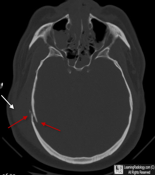
Depressed Skull Fracture. CT of the brain at bone window settings show a fracture of the right parietal bone (red arrows) with one portion of the fracture depressed beneath the inner table of the other fragment. Associated soft tissue swelling is seen (white arrow).
Imaging in Skull Fractures. AN Khan. eMedicine
|
|
|
