|
|
Calcification of the Tracheal Rings
General Considerations
- Benign mature osteogenic lesions
- Arise from membranous bones in the skull and face
- Highest incidence in 6th decade
- Female to male ratio of 3:1
- Usually involve the frontal bone
Clinical Findings
- Asymptomatic
- Slow-growing, painless mass
Imaging Findings
- Rounded, sclerotic lesions usually arising from the outer table
- Their borders are usually smooth
- The underlying cortex is not involved
- “Mature osteomas” may consist of a radiolucent nidus surrounded by dense sclerosis (ivory osteoma)
- They have no Haversian canals and no fibrous component
- Trabecular osteomas are composed of cancellous bone surrounded by denser cortex
- Gardner Syndrome is multiple skull, sinus or mandible osteomas associated with colon polyps and soft tissue skin tumors
Treatment
- Not needed unless for cosmetic reasons or from obstruction of a sinus producing mucocoele formation
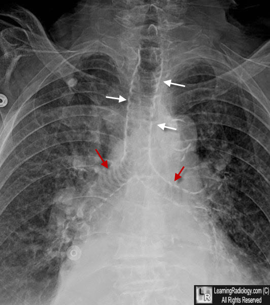
Tracheobronchial Calcification. The cartilaginous rings of the trachea (white arrows) and mainstem bronchi (white arrows) are diffusely calcified in this 95 year-old patient.
|
|
|
