|
|
Subcutaneous Emphysema
General Considerations
- Subcutaneous emphysema refers to the presence of air in the loose subcutaneous areolar tissue and muscle
- Uncommon finding
- Can occur secondary to
- Pneumomediastinum
- Pneumomediastinum occurs due to intrapulmonary rupture of alveoli and spread of air along the vascular sheath to the mediastinum
- Air spreads through loose areolar tissue and can enter the neck and subcutaneous tissues leading to subcutaneous emphysema
- Necrosis of subcutaneous tissue by gas-forming organisms (gas gangrene)
- An “air leak” in which a chest tube connected to suction inadvertently directs air into the subcutaneous tissue
- Air in subcutaneous tissue can spread in all directions
- Commonly upper parts of the body are involved more than lower body parts
- Rarely subcutaneous emphysema can occur in absence of pneumomediastinum or pneumothorax
Clinical Findings
- Subcutaneous emphysema can often produce what appears to be smooth swelling of the skin which is associated with a crunchy sensation on palpation
- Palpation produces crepitus, an unusual crackling sensation as the gas is pushed through the tissue
Imaging Findings
- On imaging studies, subcutaneous emphysema produces a striking picture of air beneath the skin surface, usually covering a large area of the body
- The air may interdigitate with the muscle bundles to produce a characteristic linear streaky pattern, especially in the pectoralis muscles over the chest
- When the streaky linear densities become widespread over the anterior and posterior chest walls, subcutaneous emphysema can obscure underlying lung pathology
- It may be impossible to detect the pleural white line of a pneumothorax
Treatment
- Treatment is to remove the cause, such as removing or repositioning the chest tube
- Air can resolve in a matter of days
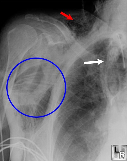
Subcutaneous Emphysema. Close-up
view of Right
Upper Lobe of
Lung and Right
Shoulder demonstrates
streaky
lucencies
overlying the
shoulder and
upper chest (blue circle) characteristic
of
subcutaneous
emphysema with
muscle bundles
of pectoralis
muscle
becoming
visible. The red arrow points to
subcutaneous
emphysema in
the
supraclavicular
area. The white arrow points to
streaky air
visible in the
mediastinum (pneumomediastinum)
Click here for
this photo
without
annotations
|
|
|
