|
|
S Sign of Golden
Reverse S Sign of Golden
General Considerations
- S Sign of Golden (AKA Golden’s S Sign, Reverse S Sign of Golden)
- The shape seen on a frontal radiograph of the chest (and CT) that consists of a mass, typically in the right hilum with associated opacification of the right upper lobe sharply demarcated by the elevated minor fissure
- The edge produced by the mass and the elevated fissure forms an “S” or “reverse S.”
- Its significance is its association with a central bronchogenic carcinoma producing the mass and the distal obstructive atelectasis - it is an ominous sign
- The sign is named after radiologist Ross Golden
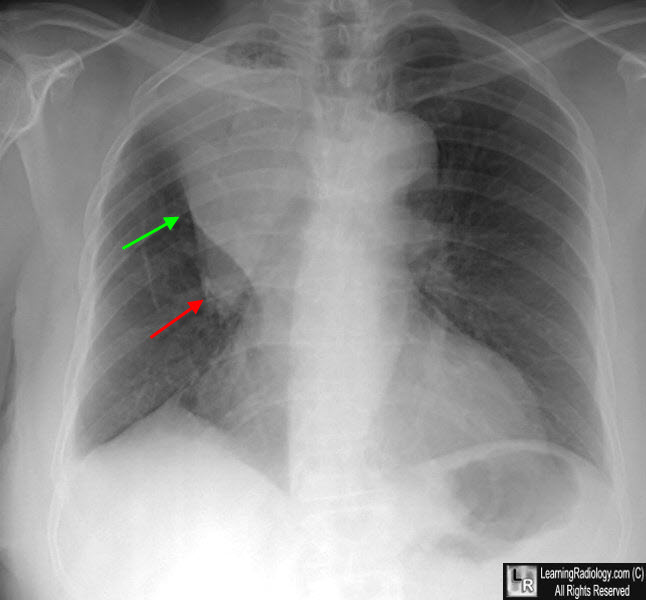
S Sign of Golden. The right upper lobe is atelectatic so the minor fissure is pulled upwards (green arrow) forming part of the "S." There is a mass in the right hilum (red arrow) which is the bronchogenic carcinoma that is causing the obstructive right upper lobe atelectasis and is forming another part of the "S."
|
|
|
