|
|
Split Pleura Sign
Empyema
General Considerations
- Enhancement of both the visceral and parietal layers of pleurae due to coating of each by fibrin resulting in capillary ingrowth
- Helps to differentiate lung abscess from empyema (pus in the pleural cavity)
Imaging Findings
- Both layers of the pleura are separated, thickened and visible
- Seen on both contrast-enhanced and non-enhanced studies (higher percentage in contrast-enhanced)
- They join at the margins of the collection
- In addition, lung abscesses have a tendency to be round whereas empyemas are lenticular in shape
- Lung abscesses tend to have a thicker and more irregularly marginated wall
Differential Diagnosis
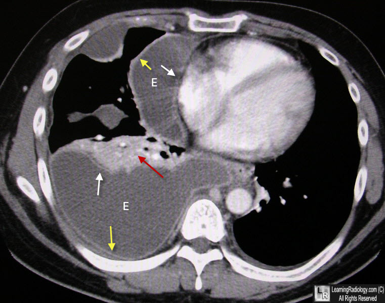
Split Pleura Sign of Empyema. At least two empyemas (E) are seen in the right hemithorax. The empyemas insinuate themselves between the visceral (white arrows) and parietal (yellow arrows) pleurae. There is associated compression atelectasis for the largest empyema(red arrow).
Differentiating Lung Abscess and Empyema: Radiography and Computed Tomography. DD Stark, MP. Federle, PC Goodman, AE. Podrasky, WR Webb. AJR 141:163-167, July 1983
|
|
|
