|
|
Spine Sign
General Considerations
- Normally, the thoracic spine “appears” to become darker (blacker) as you view it from the top to bottom on a lateral chest radiographic
- This is because more of the x-ray beam is able to penetrate the lower spine, attenuated mostly by only air in the lungs, than in the upper spine, where the beam is attenuated by the bones and muscles of the shoulder girdle
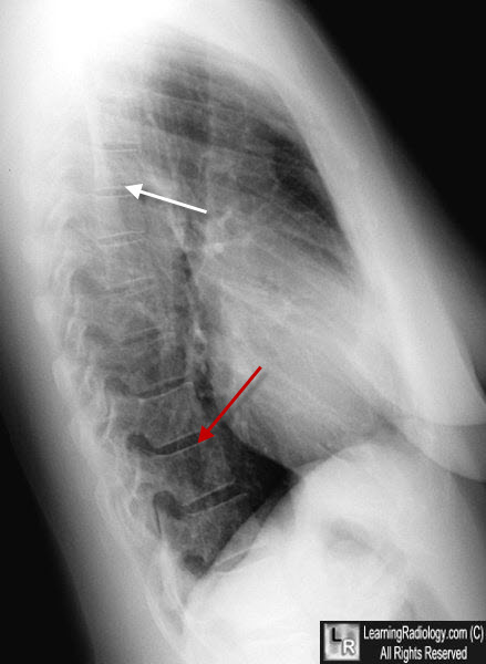
Normal Lateral Chest. On the lateral view, the thoracic spine appears to become darker (red arrow) as it approaches the hemidiaphragm because the x-ray beam traverses less dense structures than it does in the upper spine where the shoulder structures superimpose on it (white arrow).
- When something abnormal interposes itself and superimposes on the lower thoracic spine, the spine may “appear” to become whiter rather than darker just above the diaphragm
- This effect is called the “Spine Sign”
- The spine sign may indicate anything from pneumonia, a lung mass, a mediastinal mass, pleural fluid or anything of sufficient density to attenuate more x-rays passing through the lower thoracic spine than normally
- It may be particularly useful in diagnosing left lower lobe pneumonia which falls below the highest part of the left hemidiaphragm and thus may not be apparent on the frontal chest radiograph
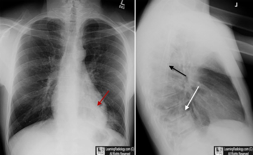
Spine Sign. On the lateral view, the thoracic spine appears whiter just above the hemidiaphragm (white arrow), the opposite of normal. The thoracic spine normally appears whiter in the region of the overlying shoulder girdle (black arrow). The left lower lobe causing this sign is also seen on the frontal radiograph, mostly hidden by the heart (red arrow).
|
|
|
