|
Silicosis
· Occupational Exposure
o Free crystalline silica (quartz) or silicon dioxide from
§ Mining
of coal, graphite, iron
§ Tin, Uranium, Gold
§ Silver, Copper
§ Also, sand blasters
§ Iron and steel foundry workers
§ Ceramic workers
§ Tunneling
· Silicosis pathophysiology
o Silica particles ingested by alveolar macrophages
o Breakdown of macrophage releases enzymes which produce fibrogenic
response
· Silicosis natural history
o Requires 10-20 years exposure before x-ray appearance
o Radiographs frequently overestimate degree of symptoms early
o Silicosis has a progressive nature despite cessation of dust exposure
· Imaging findings
o Multiple small rounded opacities 1-10 mm in size
o Usually in upper lobes
§ Mostly in apical and posterior regions of upper lobes
and apical portion of lower lobes
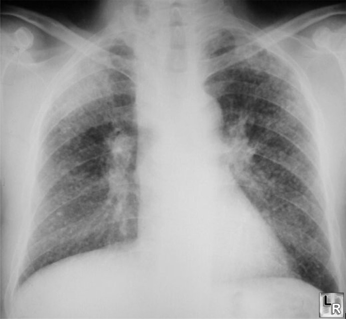
Silicosis features a diffuse micronodular lung disease
with an upper lobe predominance
o May have ground-glass appearance
o May occasionally calcify centrally (20%)
o Lymph node enlargement common
§ Eggshell calcification of hilar nodes (5%)
· DDx:
Sarcoidosis
o Large opacities are conglomerations of small opacities
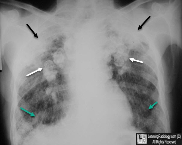
Silicosis with Progressive Massive Fibrosis. There are large conglomerate upper lobe "masses" (black arrows). Multiple enlarged and calcified hilar lymph nodes are seen, many with rim-like or "egg-shell" calcification (white arrows). There is scarring in both lower lobes (green arrows).
· Complicated Silicosis (Progressive Massive Fibrosis—PMF)
o Massive fibrosis and conglomerate nodule formation in upper lobes with
scarring and retraction of hila upwards
o Conglomerate nodules are >1 cm in size
§ Usually in mid-zone or periphery of upper lobes
§ Compensatory emphysema occurs in lower lung fields
§ Nodules tend to disappear from rest of lung when PMF
develops
o Progressive Massive Fibrosis (PMF) may cavitate from tuberculosis or
ischemic necrosis
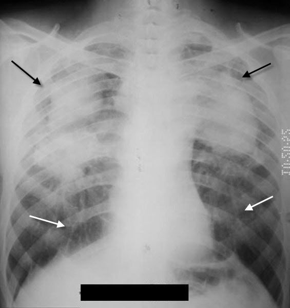
Progressive Massive Fibrosis. There are conglomerate soft-tissue densities in both upper lobes (black arrows) with linear scarring leading from the lower lobes (white arrows).
· Acute silicosis (silicoproteinosis)
o From exposure to high concentrations of silica dust
o Alveoli are filled with lipid-rich, PAS-positive
material
o Bilateral air-space disease with perihilar distribution
§ Imaging findings are similar to alveolar proteinosis
· Caplan’s Syndrome
o Consists of large necrobiotic nodules (rheumatoid nodules)
superimposed on silicosis or coal worker’s pneumoconiosis (CWP)
§ More common with CWP
o Other connective tissue diseases associated with silicosis
§ Scleroderma, RA, SLE
· Silicosis Complications
o Predisposes to TB
o Exhibits “limited” evidence for carcinogenesis in humans
|
