|
Sarcoid
General
·
Widespread formation
of non-caseating granulomas
·
3:1 female:male and
14:1 black:white predominance
·
ACE (angiotensin
converting enzyme) elevated in 70%
·
Kveim skin test
·
Positive in (70%)
but rarely used today
·
Lofgren Syndrome:
·
Acute bilateral hilar adenopathy, fever, erythema nodosum and arthralgia
·
Intrathoracic
disease (90%)
Stage I
·
Adenopathy alone
(43%)-Stage 1
·
Intrathoracic adenopathy in 80%
·
Location
·
Bilateral hilar and (R)
paratracheal
·
Most common (75-90%)
·
“1-2-3 sign”, “Pawn-broker’s sign”, “Garland sign”
·
Unilateral hilar nodes rare (3-8%)
·
Egg-shell
calcification hilar nodes in long-standing sarcoid
·
Rare
·
DDX: Silicosis
Stage II
·
Adenopathy and
parenchymal disease (41%)-Stage 2
·
Adenopathy usually
decreases as parenchymal disease increases
·
About 1/3 of
patients with adenopathy develop parenchymal disease
Stage III
·
Parenchymal disease
alone (30%)
·
Adenopathy does not
develop subsequent to parenchymal disease
·
If adenopathy develops, think of lymphoma or TB
Patterns of lung disease
·
Reticulonodular
(46%)
·
Acinar pattern (20%)
·
Larger nodules
·
“Alveolar sarcoid”
(2%)
· Coalescence of numerous interstitial granulomas
·
Air bronchograms present
·
DDX: Alveolar cell ca, alveolar proteinosis, lymphoma
Stage IV
·
End-stage lung
disease-Stage 4
·
Diffuse fibrosis
·
Bronchiectasis-honeycomb
lung
·
Multiple cysts
Uncommon manifestations
·
Associated with TB
(13%)
·
Pleural effusion
(2%)
·
Usually exudate with lymphocytic predominance
·
Cavitation of
nodules (<1%)
·
Fungus ball
formation in chronic sarcoid cavities (usually TB)
·
Focal pleural
thickening
Uncommon manifestations
·
Bronchostenosis with
lobar atelectasis
·
Pulmonary arterial
hypertension
·
Cor pulmonale
·
Pneumothorax 2°
chronic lung disease
Extrathoracic disease
·
Peripheral
adenopathy (30%)
·
Liver
·
Hepatomegaly
·
Spleen
·
Splenomegaly
·
Bone-especially
hands
·
Skin
·
Erythema nodosum
·
Lupus pernio (raised purplish nodules)
·
Muscle
·
Myopathy
·
Eyes
·
Uveitis
·
Uveoparotid fever
·
CNS
·
Granulomatous meningitis
·
Facial nerve palsy
·
Myocardium
·
Arrhythmias
·
Heart block
·
Cardiomyopathy
·
Salivary gland
·
Parotid enlargement
Prognosis
·
3/4 show complete
resolution of hilar adenopathy
·
1/3 show complete
resolution of parenchymal disease
·
20% have
irreversible pulmonary fibrosis
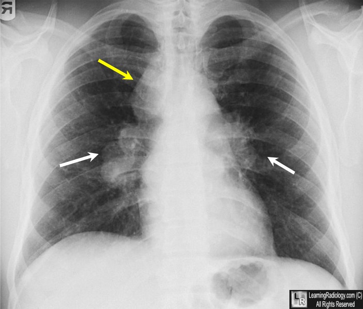
Sarcoid. There is bilateral hilar (white arrows) and right paratracheal (yellow arrow) adenopathy, the classical triad of adenopathy in pulmonary sarcoid. Notice how adenopathy produces a lobulated, lumpy contour.
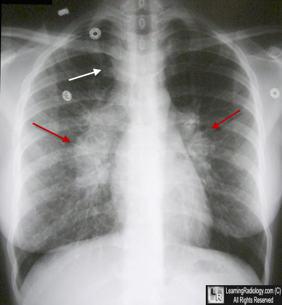
Sarcoid. There is bilateral hilar (red arrows) and right paratracheal (white arrow) adenopathy, again the classical triad of adenopathy in pulmonary sarcoid. Notice again how adenopathy produces a lobulated, lumpy contour.
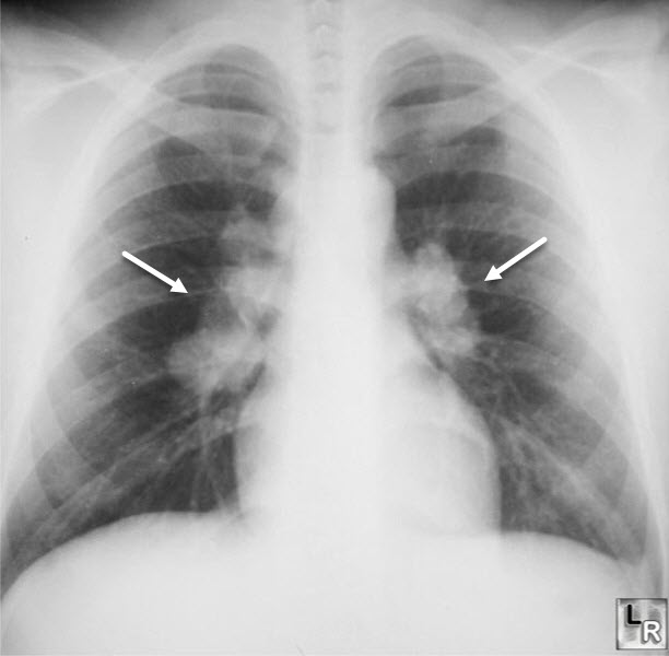
Sarcoid. There are bilaterally enlarged hilar lymph nodes (white arrows) with a clear-space noted between the nodes and the cardiac contours.
|
