|
|
Saber Sheath Deformity of the Trachea
General Considerations
- A condition in which the anteroposterior diameter of the intrathoracic trachea (sagittal diameter) exceeds the lateral diameter (coronal diameter) by a ratio of 2:1
- Strong association with chronic obstructive pulmonary disease
- Extrathoracic trachea is normal
- More common in males
Imaging Findings
- Anteroposterior elongation of the tracheal diameter and narrowing of the lateral (side-to-side) diameter by 2:1
- Slight thickening of the tracheal wall
- Inward bowing of the lateral walls which may become worse on expiration
- Ossification of the tracheal rings (which can also occur with aging)
Differential Diagnosis
- Constricting mediastinal mass
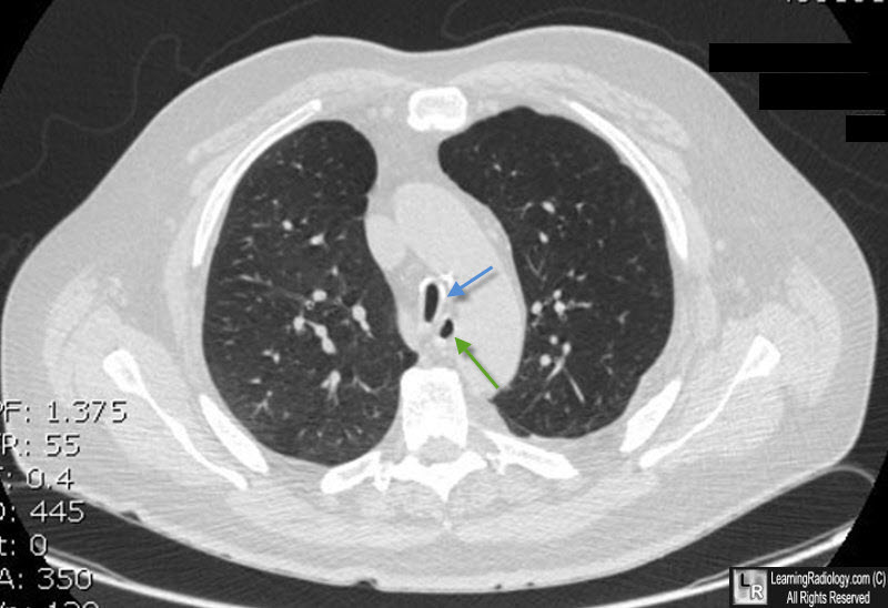
Saber Sheath Deformity of the Trachea. There is marked narrowing of the coronal (lateral) diameter of the trachea (blue arrow) and elongation of the anteroposterior (sagittal) diameter by a ratio that well exceeds 2:1, characteristic of a saber sheath trachea. The esophagus is shown by the green arrow.
“Saber-Sheath” Trachea: Relation to Chronic Obstructive Pulmonary Disease. R Greene. Am J Roentgenol 130:441-445, March 1978.
Using CT to Diagnose Nonneoplastic Tracheal Abnormalities Appearance of the Tracheal Wall. EM Webb, BM Elicker and WR Webb. May 2000, Volume 174, Number 5.
|
|
|
