|
|
Pectus Excavatum Deformity
General Considerations
- Congenital deformity of the sternum and 4 to 5 anterior ribs resulting in a funnel-shaped or sunken anterior chest wall
- Most common chest wall abnormality
- Male:female ratio 3:1
- Most cases are noticed at birth and 90% by age 1 year
- Familial in 35%
- Also associated with Marfan and Poland Syndromes
Clinical Findings
- Most are asymptomatic
- May be associated with mitral valve prolapse in 20-60%
- Pulmonary function tests can show a restrictive pattern
Imaging Findings
- Posterior displacement of the lower sternum
- Downward angulation of the anterior ribs
- Apparent cardiomegaly caused by compression of the heart
- Absence of the normal right atrial shadow to the right of the lower thoracic spine
Treatment
- A ratio of dividing the transverse diameter of the chest by the anteroposterior diameter of more than 3.2 (the Haller Index) may be an indication for corrective surgery, now often performed laparoscopically and utilizing minimally invasive surgery
- More precise chest measurements can be obtained with CT scanning than conventional radiography
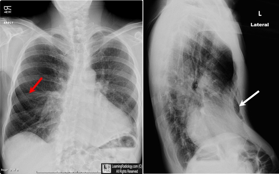
Pectus Excavatum Deformity. On the frontal projection, there is an accentuated, downward orientation of the lower anterior ribs (red arrow). The heart appears enlarged but part of that appearance is the result of compression of the heart, seen better on the lateral view. The right heart border is indistinct. On he lateral view, the lower sternum is depressed inward (white arrow), compressing the heart between it and the spine.
Pectus Excavatum Andre Hebra, MD eMedicine
|
|
|
