|
|
Mediastinal Lipomatosis
General Considerations
- Relatively common cause of mediastinal widening due to increased deposition of unencapsulated normal fat in the mediastinum
- Benign condition
- Most often associated with
- Iatrogenic steroid administration
- Cushing’s syndrome
- Simple obesity
Clinical Findings
- Usually benign and asymptomatic
- Has been reported to cause respiratory distress
- Rarely associated with tracheal compression
- Laryngeal compression
- Vena caval compression
- Regresses as steroids are tapered
Imaging Findings
- Definitive diagnosis is made by CT as mediastinal lipomatosis may mimic a mass such as
- Lymphoma
- Small cell carcinoma of the lung
- Mediastinal hemorrhage
- There are sufficient findings that, combined with history, make the findings on a chest radiograph highly suggestive
- Smooth widening of the anterior and superior mediastinum without any deformity of the trachea
- Mediastinal widening is usually bilateral and frequently symmetrical
- Contours of the widening are smooth and sharply defined
- Density may not as pronounced as those of other masses since it composed of fat
- Other fat pads in the chest may enlarge as well

- Epicardial fat pad
- Fat pad in the right cardiophrenic angle

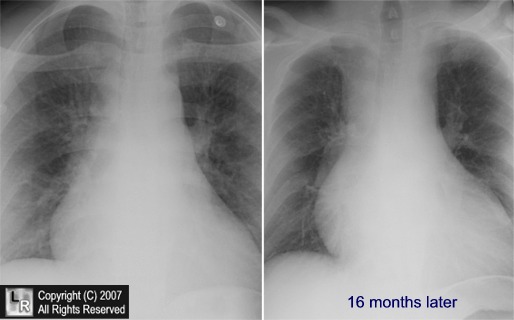
Mediastinal lipomatosis. Two frontal radiographs of the mediastinum taken 16 months apart after the patient had been taking exogenous steroids for asthma show smooth and symmetrical widening of the mediastinum (white arrows) due to increased deposition of fat in the mediastinum.
For additional information about this disease, click on this icon if above.
For this same photo without the arrows, click here
Mediastinal lipomatosis. CT confirmation of a normal variant. Homer MJ,
Wechsler RJ and Carter BL. Radiology 1978
|
|
|

)