|
|
Lung Hernia
General Considerations
- Rare
- Protrusion of lung covered by both parietal and visceral pleurae through an abnormal opening in the thoracic wall
- Most are acquired and traumatic
- Usually due to motor vehicle accident, prior chest tube insertion or coughing and rib fracture
- Other causes
- Congenital, due to developmental defects in the chest wall
- Most frequently in the supraclavicular fossa or the anterior chest wall
- In adults, cervical lung hernias are acquired, especially in older persons with chronic coughs and/or emphysema or in weight-lifters or those who play wind instruments
- Care must be exercised in inserting a central venous catheter in such people
- Secondary to infectious (e.g., TB) or neoplastic disease of the chest wall
- Post-traumatic intercostal hernias may not appear for months or years after the trauma
Clinical Findings
- Most are asymptomatic
- Palpable, soft, crepitant intercostal mass which may expand in size on inspiration or with Valsalva
- CT is more helpful in showing the origin and size of the hernia
Imaging Findings
- Lung tissue protruding outside of the thoracic cage
- May be missed on radiographs unless beam is tangential to the hernia
Treatment
- Congenital cervical hernias almost always resolve spontaneously
- Usually conservative treatment is sufficient
- If the hernia is large or lung becomes strangulated, surgery may be required
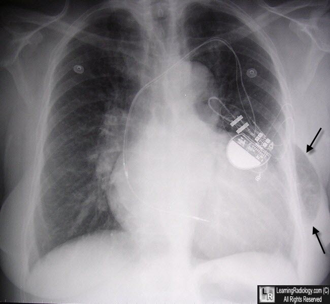
Lung Hernia. There is protrusion of lung outside the thoracic cage (black arrows), This is an acquired, post-traumatic lesion caused by the previous insertion of a chest tube.
Hernia of the Lung Through the Anterior Chest Wall. M Shameema, T Saada, R Bhargavaa, Z Ahmeda, DK Pandeya, and N
Lung Hernia: Radiographic Features. M Bhalla, BS Leitman, C Forcade, E Stern, DP Naidich and DI McCauley. AJR. Jan 1990, Vol 154, No 1. 51-53.
|
|
|
