|
|
Saline Breast Implant Calcification
General Considerations
- Calcification can occur in the fibrous capsule that develops around a breast implant
- More commonly occurs with first generation silicone implants, when the implant has been present for more than a decade, rupture of the implant
- Subglandular implants are more likely to calcify than submuscular implants
Clinical Findings
- Calcifications are considered of no clinical significance unless they mimic or obscure calcifications of malignancy on mammograms
- Implants are ovoid in shape initially but may become rounder with calcification
- May produce firmness
- May produce pain
Imaging Findings
- Calcification may be focal or diffuse
- Thin-rim of calcification that outlines implant
- Does not imply rupture
- MRI is the most accurate imaging examination for the evaluation of silicone breast implant rupture
Treatment
- None required for the calcification itself
Consumer information about breast implants is available from the FDA at this link


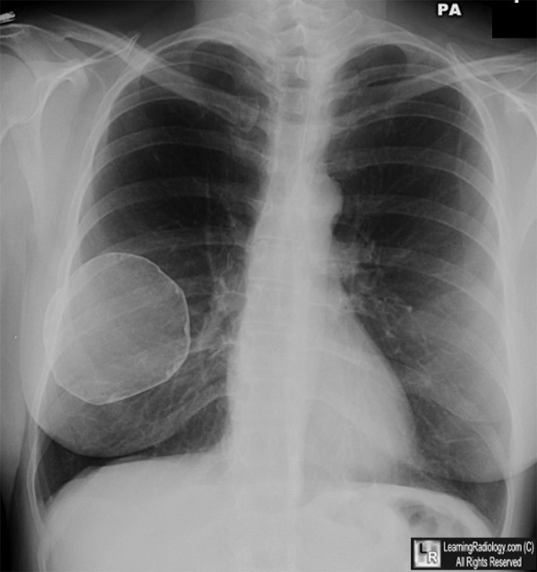
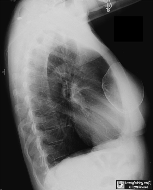
Calcified Breast Implant. Thin, curvilinear calcification (white arrows) surrounds the capsule of this patient's right saline breast implant done inserted 12 years earlier. The left implant is denser than normal breast tissue (yellow arrow), but its calcification is not visible on this chest radiograph.
For these same photos without the arrows, click here and here
For more information, click on the link if you see this icon 
|
|
|
