|
|
Epicardial (Pericardial) Fat Pad
General Considerations
- An accumulation of fat between the parietal pericardium and the parietal pleura, usually found incidentally on chest radiography
- Most common in either the right cardiophrenic angle or adherent to the left ventricle at the apex of the heart
- On the left side, it blunts the normal rounded apex of the heart
- Can be mistaken for pneumonia or a mass
- May or may not be associated with visceral obesity
- Some studies show an association between increased epicardial fat and cardiac abnormalities, such as coronary artery disease
- May also become larger in mediastinal lipomatosis caused by Cushing syndrome or exogenous steroid administration
Clinical Findings
Imaging Findings
- “Mass” of fat density that does not silhouette the adjacent soft tissue of the heart or diaphragm, usually seen best on frontal radiographs
- Most frequently seen at the left cardiophrenic angle and then at the right cardiophrenic angle
- Remains stable over serial images
- Low Hounsfield (negative) numbers on CT
Differential Diagnosis
- Morgagni hernia

- Pericardial cyst
- The epicardial fat pad described here is a normal finding
- The epicardial fat pad sign (pericardial fat pad sign, fat pad sign) is an abnormal finding that can be seen with pericardial effusion
- Curvilinear fat density in displaced posteriorly from sternum on lateral chest radiograph
Treatment
- Incidental finding requiring no treatment


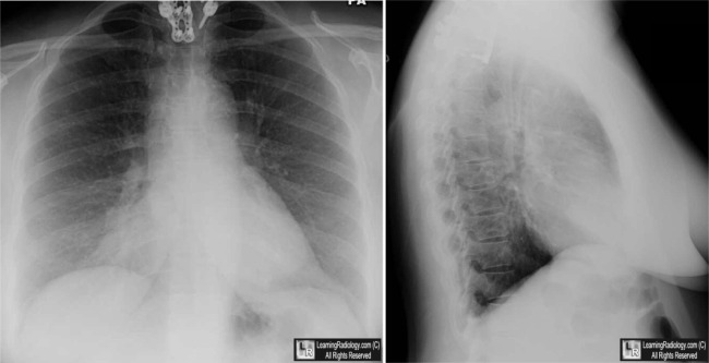
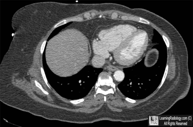
Epicardial (Pericardial) Fat Pad. Above: Frontal and lateral radiographs of the chest show an area of increased density at the right cardiophrenic angle on the frontal radiograph (white arrow) that does not silhouette the heart border (black arrow).There is an area of slightly increased density suggested on the lateral view (white arrow). Below: An axial CT scan through the lower thorax shows an accumulation of fat (yellow arrow) in the space anterior to the heart and the right hemidiaphragm (D).
For this same photo without the arrows, click here and here
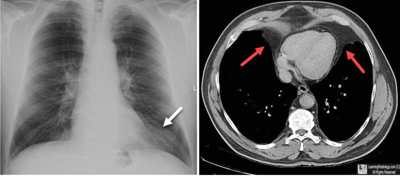
Epicardial (Pericardial) Fat Pad. Another case: Frontal chest radiograph shows an area of increased density at the left cardiophrenic angle (white arrow) that does not silhouette the heart border. An axial CT scan through the lower thorax shows an accumulation of fat in both the left and right cardiophrenic angles (red arrows).
For more information, click on the link if you see this icon 
Epicardial Fat and Its Association with Cardiovascular Risk: A Cross-Sectional Observational Study. F Mookadam, R Goel, MS Alharthi, P Jiamsripong, and S Cha. Heart Views. 2010 Oct-Dec; 11(3): 103–108.
The Right Pericardial Fat Pad. SL Cohen. March 1953 Radiology, 60, 391-393.
|
|
|
