|
|
Eggshell Calcification
General Considerations
- Calcification around the periphery of lymph nodes
- Reason for this pattern of calcification is not known
- Most commonly seen in silicosis and coal-worker's pneumoconiosis (3-6%)
- Also seen in sarcoid (5%)
- Rarely in post-irradiated Hodgkin disease, blastomycosis, scleroderma, amyloid and histoplasmosis
Imaging Findings
- Established criteria include
- Calcification <2mm thick in the periphery of a lymph node
- Ring-like shadow must be complete in at least one of the nodes involved
- One of the nodes must be at least 1 cm in size
- There may or may not be interior calcifications
Differential Diagnosis
- Pulmonary artery aneurysms
- Calcification of pulmonary arteries from prolonged pulmonary arterial hypertension
 
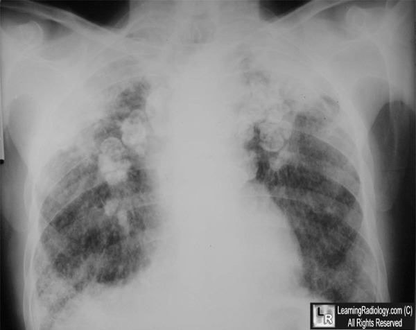
Eggshell Calcifications in Silicosis. The white arrows point to enlarged hilar lymph nodes with peripheral "eggshell" calcifications characteristic of either silicosis or sarcoidosis. This patient also has upper
lobe fibrosis seen with silicosis and had a history of being a sandblaster.
For this same photo without the annotations, click here or here
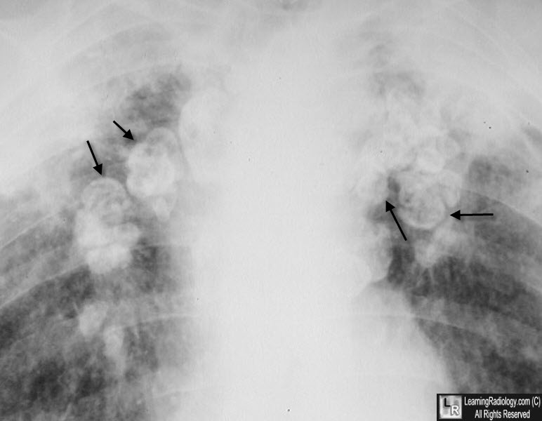
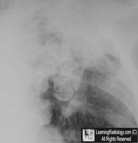
Eggshell Calcifications in Silicosis. Close-up of the same patient as above. The black arrows point to enlarged hilar lymph nodes with peripheral "eggshell" calcifications characteristic of either silicosis or sarcoidosis.
Eggshell Calcification of Lymph Nodes. Gross, B; Schneider, HJ and Proto, A. AJR:135, December 1980
|
|
|
