|
|
Left Upper Lobe Atelectasis
Patterns of Collapse in Lobar Atelectasis
- Obstructive atelectasis produces consistently recognizable patterns of collapse depending on the location of the atelectatic segment or lobe and the degree to which such factors as collateral airflow between lobes and obstructive pneumonia allow the affected lobe to collapse.
- In general, lobes collapse in a fan-like configuration with the base of the fan-shaped triangle anchored at the pleural surface and the apex of the triangle anchored at the hilum.
- Other, unaffected lobes will undergo compensatory hyperinflation in an attempt to “fill” the affected hemithorax and this hyperinflation may limit the amount of shift of the mobile chest structures.
Left upper lobe atelectasis
- On the frontal radiograph:
- There is a hazy area of increased density around the left hilum.
- There is a leftward shift of the trachea.
- There may be elevation with "tenting" (peaking) of the left hemidiaphragm.
- Compensatory overinflation of the lower lobe may cause the superior segment of the left lower lobe to extend to the apex of the thorax on the affected side.
- On the lateral radiograph:
- There is forward displacement of the major fissure and the opacified upper lobe forms a band of increased density running roughly parallel to the sternum.
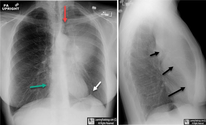
Left Upper Lobe Atelectasis. On the frontal view, there is a hazy density surrounding the left hilum. The heart is displaced towards the left (green arrow). There is "tenting" of the left hemidiaphragm (white arrow). A portion of the left lower lobe expands superior and medial to the collapsed lower lobe (red arrow) ("Luftsichel sign"). On the lateral view, the major fissure (black arrows) is displaced forward due to volume loss in the left upper lobe.
|
|
|
